

For decades, radiologists, orthopedic surgeons and rheumatologists have relied on a set of X-ray images to fully assess a patient’s osteoarticular condition. AP and LAT views have been used to help physicians imagine what a 3D skeletal image would look like.
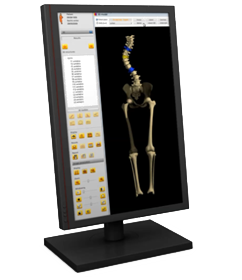
Now, using only two EOS® low dose images, the sterEOS® workstation enables personalized 3D bone modeling of the patient in a natural weight-bearing position (spine and/or lower limbs) and automatic calculation of clinical parameters.
EOS® 3D images are created via patented sterEOS® software from two unique low dose images, with no additional radiation. The algorithms are based on the use of statistical modeling and bone shape recognition. This modeling falicitates the automatic calculation of many tridimensional clinical parameters.
3D skeletal envelope images can be obtained for the spine, the femur and the tibia. Unlike CT, EOS® images are acquired in an upright position, enabling new ways to globally evaluate a patient's postural abnormality. SterEOS® 3D modeling allows the display of bone position, rotation and orientation. It also enables the display in different perspectives, in conjunction with the calculation of over 100 clinical parameters relevant for surgical planning and follow-up of pathologies.
Clinical benefits
- Visualize and understand the position, rotation and orientation of the bones in 3D and within the global skeletal system from a weight-bearing position
- Improve diagnosis and clinical research capabilities with over 100+ automatically calculated, accurate 2D and 3D measurements6that are free from bias and taken in a functional position
- Perform a post-operative assessment of a total hip replacement through a dedicated and precise workflow that automatically calculates the position of the prosthetic components
- Visualize 3D models and calculate 2D/3D clinical parameters even from pediatric Micro Dose exams and seated exams with the EOS Radiolucent Chair
Facility-wide efficiency
- Simple, straightforward 3D workflows dedicated to postural assessment, spine, lower limbs and Total Hip Arthroplasty (THA)
- Powerful patient database with user access management for online and off-line retrieval by referring physicians
- Seamless push/pull on PACS and webPACS of images, patient reports and 3D models
- Direct link to the online EOS 3DServices and to the EOSapps, 3D surgical planning solutions
Bibliography
- Comparison of radiation dose, workflow, patient comfort and financial break-even of standard digital radiography and a novel biplanar low-dose X-ray system for upright full-length lower limb and whole spine radiography. Dietrich TJ et al. Skeletal Radiol. 2013.
- Diagnostic imaging of Spinal deformities: Reducing Patients Radiation Dose With a New Slot-Scanning X-ray Imager. Deschenes S, Charron G, Beaudoin G,Labelle H, Dubois J, Miron M, Parent S. Spine April 2010, 35 (9): 989. .
- Ionizing radiation doses during lower limb torsion and anteversion measurements by EOS stereoradiography and computed tomography. Delin C et al. Eur J Radiol. 2014
- EOS microdose protocol for the radiological follow-up of adolescent idiopathic scoliosis. Ilharreborde B. et al. Eur Spine J. 2015
- Preoperative three-dimensional planning of total hip arthroplasty based on biplanar low-dose radiographs: accuracy and reproducibility for a set of 31 patients. Mainard, D et al. Communication at ISTA 2014.
- The EOS imaging system and its uses in daily orthopaedic practice. Illes T, Somoskeoy S. Int Orthop2012 Feb 28.
- Accuracy of Digital Preoperative Templating in 100 Consecutive Uncemented Total Hip Arthroplasties, Journal of Arthroplasty, 2013-02-01, R.Shaarani & al
- What proportion of patients report long-term pain after total hip or knee replacement for osteoarthritis? A systematic review of prospective studies in unselected patients. Beswick, A. D., V. Wylde, R. Gooberman-Hill, A. Blom and P. Dieppe (2012). BMJ Open.
- Meijjo Hospital, Nagoya, Japan
More products from this supplier
- United States
- United States
- United States
- United States
- United States
- United States
- United States
Loading...


