
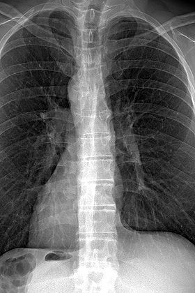
The EOS system provides low dose, full body, stereo-radiographic images of your patient in a functional position. The EOS system is designed around a vertically traveling arm supporting two image acquisition systems mounted at right angles. Each acquisition system is composed of an X-ray tube and a linear detector.
This unique biplanar design and linear, vertical scanning technique acquires frontal and lateral images of your patient simultaneously in either a standing or seated position with the EOS Radiolucent Chair.
Clinical benefits
- Comprehend compensation mechanisms between the spine, hip and knee thanks to full body, weight-bearing images
- Calculate precise 2D and 3D measurements, free from magnification and stitching bias
- Improved diagnostics due to high image quality and over 65,000 grey levels for excellent contrast
Dose reduction
- Patient’s radiation dose decreased by 50% compared to a DR system1and 85% compared to a CR system2
- Substitution of specific CT exams with an EOS exam to reduce the patient’s radiation dose by 95%3
- Micro Dose protocol for a full spine exam (frontal and lateral) at a dose that’s equivalent to only a week’s worth of natural radiation4
Facility-wide efficiency
- Capture frontal and lateral, full body images in less than 20 seconds for adults and 15 seconds for children
- Complete an exam in under 4 minutes, even for complex spine or full body1
- Maximize patient throughput with up to 120 complex exams per day9
- Facilitate the process to image disabled patients with the EOS Radiolucent Chair
Bibliography
- Comparison of radiation dose, workflow, patient comfort and financial break-even of standard digital radiography and a novel biplanar low-dose X-ray system for upright full-length lower limb and whole spine radiography. Dietrich TJ et al. Skeletal Radiol. 2013.
- Diagnostic imaging of Spinal deformities: Reducing Patients Radiation Dose With a New Slot-Scanning X-ray Imager. Deschenes S, Charron G, Beaudoin G,Labelle H, Dubois J, Miron M, Parent S. Spine April 2010, 35 (9): 989. .
- Ionizing radiation doses during lower limb torsion and anteversion measurements by EOS stereoradiography and computed tomography. Delin C et al. Eur J Radiol. 2014
- EOS microdose protocol for the radiological follow-up of adolescent idiopathic scoliosis. Ilharreborde B. et al. Eur Spine J. 2015
- Preoperative three-dimensional planning of total hip arthroplasty based on biplanar low-dose radiographs: accuracy and reproducibility for a set of 31 patients. Mainard, D et al. Communication at ISTA 2014.
- The EOS imaging system and its uses in daily orthopaedic practice. Illes T, Somoskeoy S. Int Orthop2012 Feb 28.
- Accuracy of Digital Preoperative Templating in 100 Consecutive Uncemented Total Hip Arthroplasties, Journal of Arthroplasty, 2013-02-01, R.Shaarani & al
- What proportion of patients report long-term pain after total hip or knee replacement for osteoarthritis? A systematic review of prospective studies in unselected patients. Beswick, A. D., V. Wylde, R. Gooberman-Hill, A. Blom and P. Dieppe (2012). BMJ Open.
- Meijjo Hospital, Nagoya, Japan

More products from this supplier
- United States
- United States
- United States
- United States
- United States
- United States
- United States


Loading...