Diagnostic confidence and greater efficiency with syngo.via General Engine
Software that “understands” human anatomy
Meaningful diagnostic reports with syngo.via Advanced Reporting
Siemens Healthcare introduces version VA30 of its routine 3D and advanced reading software syngo.via, featuring new applications and functionalities that further streamline and accelerate the syngo.via experience. The latest innovation is the syngo.via General Engine, a new package of highly automated and standardized applications. “Anatomical Range Presets,” for example, identifies individual regions of the body on images captured using computed tomography (CT) and magnetic resonance imaging (MRI), aligns the image projections accordingly, then selects detailed views to facilitate case preparation. For customers, this means greater efficiency and enables higher diagnostic confidence.
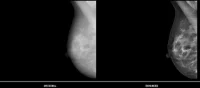
Siemens is making syngo.via more anatomically intelligent to support radiologists and medical technology personnel in their routine workflows. The software “understands” human anatomy and prepares the images for diagnostic reading. Syngo.via VA30 features “Automatic Rib Labeling,” for example, which automatically identifies and labels the ribs in CT scans. Until now, radiologists had to identify the ribs manually. Given the unique shape of the ribs, this can be time-consuming and lead to errors, especially in complex diagnostic environments such as oncology.
Anatomical intelligence is also a hallmark of the new syngo.via General Engine software package with which customers can upgrade their software. The package includes the “Anatomical Range Presets” feature, which displays a quick, precise, optimal view of selected anatomical regions. To facilitate diagnoses, users often create such views manually, performing the multi-step process of selecting the relevant area, aligning the image projections accordingly, and editing the detailed view. This takes time, requires anatomical expertise, and is error-prone. The new application from Siemens largely automates these steps. This delivers a consistent quality of the resulting anatomical views and snapshots that does not depend on the skill of the specific user. Using a technology akin to facial recognition in digital photography, syngo.via is able to recognize shoulders, spines, hips, etc. in clinical images and optimize how they are displayed in their anatomical environment. The presets are initially available for specific anatomical regions in CT and MRI images.
Following an examination, the radiological diagnostic report plays a key role in the treatment of the patient. The syngo.via Advanced Reporting tool, part of the syngo.via General Engine, helps radiologists create clear, well-structured reports for the referring or follow-up physicians. Standardized templates make it easier to create reports but can still be customized to individual needs, and findings from multiple examinations can be consolidated into a single report. In the past, multiple diagnoses meant having different documents pertaining to a single case. Now, the diagnostic report reflects the patient’s entire disease profile. This makes it much easier for doctors to form a comprehensive assessment of the patient’s condition, which helps to improve the quality of treatment.
Source: Siemens
26 November 2013
Latest Articles
Imaging, Siemens, Software
Diagnostic confidence and greater efficiency with syngo.via General Engine Software that “understands” human anatomy Meaningful diagnostic reports w...