Opportunity exists for substantial radiation dose reduction using existing computed tomography (CT) technology for common diagnostic tasks, according to a new study reported in the journal Academic Radiology.
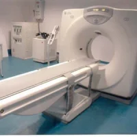
CT is widely used in medical practice and can provide images with high spatial and temporal resolution that cover large regions of the body, with few contraindications to imaging. The most notable concern for CT imaging is that it requires exposure to ionising radiation. It's important to use the optimal dose of radiation that will accomplish the diagnostic task but not compromise diagnostic performance. However, this “optimal” dose is not known for even the most common CT examinations, resulting in a wide spectrum of radiation doses being used.
The lack of available evidence on what dose levels are required to achieve specific results is due to many factors, for example, evolving technology or fear that substantial lowering of the dose may result in compromised diagnostic benefit. Several recent technical and clinical advances have expanded opportunities to lower radiation dose: automatic exposure control (AEC) and automated tube potential selection adapt the radiation dose to patient size and diagnostic task; while iterative reconstruction substantially improves the image quality of lower-dose CT images, but with an unclear impact performance for many diagnostic tasks.
In this study, researchers estimated observer performance for a range of dose levels for common CT examinations (detection of liver metastases or pulmonary nodules, and cause of neurologic deficit) to prioritise noninferior dose levels for further analysis. Using CT data from 131 examinations (abdominal CT, 44; chest CT, 44; head CT, 43), CT images corresponding to 4%–100% of the routine clinical dose were reconstructed with filtered back projection or iterative reconstruction (IR).
Radiologists evaluated CT images, marking specified targets, providing confidence scores, and grading image quality. Noninferiority was assessed using reference standards, reader agreement rules, and jackknife alternative free-response receiver operating characteristic figures of merit. Reader agreement required that a majority of readers at lower dose identify target lesions seen by the majority of readers at routine dose.
Reader agreement identified dose levels lower than 50% and 4% to have inadequate performance for detection of hepatic metastases and pulmonary nodules, respectively, but could not exclude any low dose levels for head CT. Estimated differences in jackknife alternative free-response receiver operating characteristic figures of merit between routine and lower dose configurations found that only the lowest dose configurations tested (i.e., 30%, 4%, and 10% of routine dose levels for abdominal, chest, and head CT examinations, respectively) did not meet criteria for noninferiority. At lower doses, subjective image quality declined before observer performance. IR was only beneficial when filtered back projection did not result in noninferior performance.
"For the diagnostic tasks examined, there appears to be substantial opportunity for radiation dose reduction, with dose reductions of 70% or more demonstrating noninferiority compared to current routine clinical dose levels," the authors note. "Because such a large variability exists between the doses needed to accomplish different diagnostic tasks, radiation dose should be reduced by adapting scan parameters to the diagnostic task performed by the radiologist rather than simply the body part being imaged."
Researchers say further work with more readers and cases will be required to better define lower radiation dose levels that do not degrade observer performance.
Source: Academic Radiology
Image Credit: Pixabay
References:
Fletcher, Joel G et al. (2017) Estimation of Observer Performance for Reduced Radiation Dose Levels in CT: Eliminating Reduced Dose Levels That Are Too Low Is the First Step. Academic Radiology. doi.org/10.1016/j.acra.2016.12.017
Latest Articles
Radiation, computed tomography, dose reduction, radiation exposure
Opportunity exists for substantial radiation dose reduction using existing computed tomography (CT) technology for common diagnostic tasks, according to a new study reported in the journal Academic Radiology.