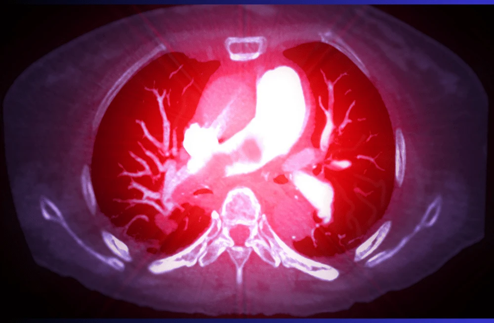
Spotting pulmonary embolism (PE) through computed tomography pulmonary angiography (CTPA) hinges on achieving sufficient contrast flow rates for clear visualization of pulmonary arteries. However, challenges arise when patients have difficult intravenous access, complicating the administration of high flow rates needed for optimal imaging.
PE protocols are crucial due to their role in diagnosing a condition that affects approximately one in every 1,000 individuals annually in the United States. The routine nature of these scans means that medical professionals frequently encounter patients with problematic IV access. Administering a bolus of contrast at high flow rates, as typically required in PE protocols, can pose a risk of vein damage or failure, further complicating patient care.
Research Approach and Findings
In a recent study published in the Journal of Medical Imaging and Radiation Sciences, researchers investigated whether a low volume, low flow rate (LVLF) CTPA protocol could offer imaging quality comparable to that of a standard protocol. The study involved 151 patients who underwent CTPA using a 320-slice multi-detector CT scanner at a hospital in Singapore. Of these, 80 patients followed the standard protocol, receiving a fixed flow rate of 4.5ml/s and 50ml of contrast media. The remaining 71 patients underwent the LVLF protocol, which included reductions of up to 37% in flow rate and 30% in contrast volume administered.
Comparative Analysis of Results
Two independent radiographers evaluated the attenuation (measured in Hounsfield units, HU) of multiple pulmonary arteries across both protocols. They found no significant differences in HU measurements for any of the seven key regions of interest—including the main pulmonary trunk, right and left pulmonary arteries, and various lobar and subsegmental arteries—between the LVLF and standard protocols. Moreover, two independent radiologists assessed overall image quality using a standardized 5-point questionnaire and reported no significant variations between the LVLF and standard protocols.
Clinical Implications for Low Contrast Volume
Lead author Wan Chin Lee, a radiographer at Changi General Hospital, and co-authors concluded that their findings support the feasibility of optimizing contrast doses and flow rates in CTPA protocols. This optimization not only maintains diagnostic efficacy but also mitigates the risks associated with high flow rates, particularly in patients with challenging IV access. The study suggests that adopting LVLF protocols could potentially lead to safer and more efficient practices in diagnosing pulmonary embolism, paving the way for broader application and further refinement in clinical settings. As medical imaging techniques advance, such research highlights the importance of balancing procedural efficacy with patient safety in diagnostic imaging protocols.
Source Credit: Journal of Medical Imaging and Radiation Sciences
Image Credit: iStock


