Retroperitoneal benign nerve sheath tumours (RBNSTs) are uncommon, comprising around 5% of retroperitoneal tumours, mainly Schwannomas and neurofibromas. Schwannomas typically occur sporadically, while neurofibromas can be solitary or associated with neurofibromatosis type 1 (NF1). Malignant transformation is rare. These tumours are often discovered incidentally during imaging for unrelated issues. Despite their benign nature, accurate identification is crucial to differentiate them from malignancies. RBNSTs lack specific radiological features, making diagnosis challenging. This review focuses on correlating histological and radiological findings to enhance diagnostic accuracy and distinguish RBNSTs from other retroperitoneal neoplasms. An educational review published in Insights into Imaging delves into the intricate aspects of RBNSTs, shedding light on their pathology, radiological features, differential diagnosis, and surgical management strategies. Investigating the correlation between tissue architecture and appearance on imaging can help increase the accuracy of differential diagnosis with other retroperitoneal neoplasms at CSIs.
Pathological Features
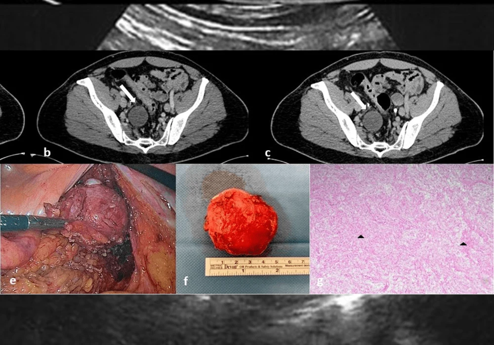
Macroscopically, RBNSTs typically manifest as solid masses with occasional cystic components. Schwannomas exhibit encapsulation and eccentric growth relative to nerve fibres, while neurofibromas lack a distinct capsule. Microscopically, Schwannomas display hypercellular "Antoni A" areas with Verocay bodies, expressing S100 and SOX10, while neurofibromas comprise Schwann cells, perineurial-like cells, and fibroblasts, often accompanied by nerve fibres and myxoid matrix.
Radiological Features
While ultrasound offers preliminary insights into RBNSTs, computed tomography (CT) and magnetic resonance imaging (MRI) serve as primary modalities for diagnosis. CT reveals hypodense lesions with minimal enhancement, while MRI showcases variable signal intensities on T1- and T2-weighted images, often exhibiting contrast enhancement. Additionally, 18F-fluorodeoxyglucose-positron emission tomography/CT (FDG-PET/CT) highlights FDG accumulation in solid tumour components.
Correlation with Pathology
MRI characteristics mirror histological composition, with myxoid-rich areas appearing hyperintense on T2-weighted images and compact areas displaying contrast enhancement. Schwannomas and neurofibromas may exhibit distinctive imaging signs such as the "target sign" or "fascicular sign", aiding in their differentiation. However, histological confirmation remains imperative for definitive diagnosis.
Differential Diagnosis
Distinguishing RBNSTs from other retroperitoneal neoplasms necessitates meticulous evaluation of location, growth pattern, and imaging features. Conditions such as carcinoma metastasis, lymphomas, and extragonadal germ cell tumours present diagnostic challenges, underscoring the importance of histopathological analysis for accurate characterisation.
Surgical Treatment
Surgical excision is the cornerstone of management for RBNSTs, with a preference for function-sparing surgery to preserve neural integrity. Minimally invasive approaches, including laparoscopy and robotics, offer safe and effective options for tumour resection. Surgical planning considers factors such as tumour size, location, and proximity to adjacent structures, ensuring optimal outcomes.
RBNSTs present a complex diagnostic and management scenario, requiring a multidisciplinary approach involving radiologists, pathologists, and surgeons. Understanding the pathological features, radiological manifestations, and surgical principles is crucial for accurate diagnosis and optimal patient outcomes.
In summary, navigating the intricate landscape of RBNSTs necessitates a nuanced understanding of their pathology, imaging characteristics, and surgical considerations, ultimately guiding tailored management strategies for each patient.
Source & Image Credit: Insights into Imaging

