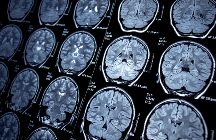
Brain metastases (BMs) are common and contribute significantly to poor patient prognosis. Local radiotherapy like stereotactic radiotherapy (SRT) and stereotactic radiosurgery (SRS) are increasingly used for BM treatment. Accurate determination of the gross tumour target volume (GTV) is crucial for their effectiveness, usually done through contrast-enhanced (CE) MRI. However, single-phase scans may not fully capture BM boundaries due to the dynamic nature of contrast agent penetration. Time-delayed CE MRI has shown promise in improving BM detection. A recent study published in Radiotherapy and Oncology explores how different delay times in CE MRI affect the visualisation, volume, and shape of large-volume BMs, aiming to refine GTV determination for better treatment planning.
Investigating Dynamic Contrast-Enhanced MRI in Brain Metastases
Between February and October 2021, the prospective study led at the Cancer Hospital Affiliated with Shandong First Medical University enrolled 188 patients with brain metastases (BMs), totalling 1,014 lesions. Patients did not receive radiotherapy (RT) or steroids. They underwent contrast-enhanced (CE) T1-weighted imaging (T1WI) at various intervals following gadolinium-based contrast agent injection. Lesions larger than 1 cm were identified from the 1-minute delayed CE T1W images, resulting in 561 lesions from 155 patients. The patients, with an average age of 62.2 years for men and 59.4 years for women, had primary tumour types, including lung, breast, and oesophagal cancers, and others. The study, approved by the Ethics Committee of Shandong Cancer Hospital, employed a Philips Brilliance Big Bore Computed Tomography (CT) locator for patient positioning and imaging. Subsequent to CT simulation, patients underwent imaging with a GE 3.0 T superconducting magnetic resonance (MR) scanner, capturing enhanced 3D T1-weighted gradient echo brain volume imaging (T1 BRAVO) at 1, 3, 5, 10, 18, and 20 minutes post-contrast injection.
Deciphering Contrast Agent Kinetics in Brain Metastases Visualisation
An in-depth analysis of patient demographics unveiled a diverse population, characterised by varying ages and primary tumour types. However, the primary focus of the study was on elucidating the dynamic changes in contrast agent concentration and their implications for GTV determination. The findings revealed a nuanced interplay between contrast agent kinetics and BM visualisation. The accumulation of contrast agents in the extracellular spaces of BMs led to pronounced enhancement in the MRI images, with both tumours and brain white matter exhibiting peak enhancement at 3 minutes post-injection. Subsequent observations indicated a gradual decline in signal intensity, highlighting the temporal dynamics of contrast agent distribution within BMs.
Optimising Radiotherapy Planning: The Role of Multi-Phase CE-MRI
Further analyses delved into the volumetric characteristics of BMs, shedding light on the differential growth rates observed between lesions of varying sizes. Interestingly, larger-volume BMs exhibited a slower growth rate compared to their smaller counterparts, attributed to inherent differences in contrast agent extravasation dynamics. Importantly, the study underscored the pivotal role of multi-phase CE-MRI scans, particularly those commencing at ≥ 10 minutes post-injection, in facilitating accurate GTV determination for large-volume BMs. This nuanced approach stands in stark contrast to conventional single-phase scans, which may fail to capture the dynamic changes in BM boundaries over time.
Clinical Implications of Enhanced GTV Determination
From a clinical standpoint, these findings have profound implications. Accurate GTV determination serves as the cornerstone of effective radiotherapy planning, with far-reaching implications for treatment efficacy and patient outcomes. Consequently, the study advocates for a paradigm shift towards the adoption of multi-phase CE-MRI scans, coupled with meticulous analysis of contrast agent kinetics, to enhance the precision of GTV delineation in large-volume BMs. This forward-looking approach promises to revolutionise radiotherapy practices, thereby improving the quality of care for patients battling BMs.
Despite the invaluable insights gleaned from this study, certain limitations warrant acknowledgement. Chief among these is the absence of histological verification, which could have provided additional validation for the observed findings. Moreover, the study's focus on scanning times limited to 20 minutes precludes a comprehensive understanding of the long-term dynamics of contrast agent distribution within BMs. Nevertheless, these limitations notwithstanding, the study represents a seminal contribution to the field, laying the groundwork for future research endeavours to unravel the complexities of BM radiotherapy planning.
Source: Radiotherapy and Oncology
Image Credit: iStock


