


Non-alcoholic fatty liver disease (NAFLD), also known as metabolic dysfunction-associated steatotic liver disease (MASLD), affects over half of the global type 2 diabetes population and is a risk factor for cardiac remodelling and heart failure. The link between hepatic fibrosis in NAFLD and increased mortality in acute heart failure, as well as left ventricular (LV) diastolic dysfunction, has been established. However, research has mainly focused on patients with advanced liver cirrhosis from tertiary centres, overlooking the progressive nature of NAFLD, which starts with steatosis, inflammation, and subclinical fibrosis before cirrhosis.
Few studies have explored the relationship between liver fibrosis and cardiac remodelling in non-cirrhotic patients, and none have used magnetic resonance imaging (MRI). MRI provides precise, non-invasive measurements of hepatic fat content and fibrosis, and detailed assessment of cardiac function and structure. A study recently published in European Radiology applied advanced cardiac and hepatic MRI techniques to investigate these relationships in a general population sample, aiming to understand the associations between liver fibrosis and cardiac structure, function, and tissue characteristics. The study also analysed circulating biomarkers of cardiovascular inflammation and fibrosis, hypothesising a significant association between liver fibrosis and LV characteristics.
Study Design and Participant Characteristics in a SCAPIS Sub-Study
The current study was a prospective cross-sectional sub-study within SCAPIS (Swedish CArdioPulmonary bioImage Study), a large-scale cohort study of over 30,000 individuals aged 50–65 from the general Swedish population. Participants were recruited from the SCAPIS cohort in Linköping, including both individuals with type 2 diabetes (T2D) and matched controls without diabetes. Exclusion criteria included type 1 diabetes, contraindications to MRI, or irregular ventricular rhythm.
Between November 2017 and July 2018, participants underwent a magnetic resonance scan at Linköping University Hospital using a 1.5 T Philips Achieva dStream scanner. The scan assessed liver fat, liver stiffness (using 3D magnetic resonance elastography, MRE), and left ventricular (LV) structure, function, and tissue characteristics. Liver stiffness was measured using an electrodynamic transducer, with results processed and reported in kPa.
The study enrolled 92 participants (mean age 59.5 years, 32 women), equally divided between those with T2D and controls. Among participants, 12% had a history of cardiac disease, and the average alcohol consumption was 45 g/week, with one participant exceeding 210 g/week. Women had higher HDL cholesterol levels than men, but no other significant sex differences were noted. The diabetes group had higher BMI, HbA1c, and alanine aminotransferase (ALT) levels compared to controls, with 21.7% of the diabetes group receiving insulin treatment.
Link Between Liver Fibrosis and Cardiac Remodelling in Non-Alcoholic Fatty Liver Disease
This study employed advanced magnetic resonance imaging (MRI) techniques to explore the relationship between liver fibrosis and cardiac remodelling in a general population sample. Most participants exhibited hepatic steatosis, with NAFLD likely being the primary cause due to the low reported alcohol consumption and the high prevalence of NAFLD in Sweden. Liver stiffness measurements did not indicate advanced fibrosis, as all values were below the cirrhosis threshold (4.1 to 4.4 kPa), with the highest value observed being 2.9 kPa. Despite the absence of advanced fibrosis, liver stiffness was independently associated with increased left ventricular (LV) concentricity, even after adjusting for liver fat and diabetes. This association was further attenuated when adjusting for circulating levels of interleukin-1 receptor type 2 (IL-1RT2), a biomarker linked to cardiac remodelling. In previous studies, IL-1RT2, a soluble decoy receptor, negatively regulates IL-1β inflammatory activity and has been associated with adverse cardiac remodelling.
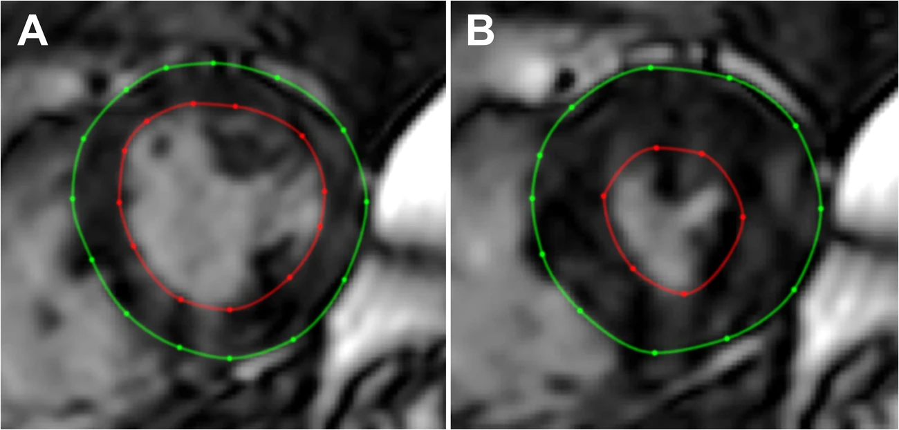
Image Credit: European Radiology
MRI Insights into Liver Stiffness and Cardiac Remodelling in Non-Cirrhotic Individuals
The term ‘liver-heart axis’ describes the impact of liver disease on heart function. In cirrhosis, patients often develop ‘cirrhotic cardiomyopathy,’ characterised by impaired systolic response to stress, resting diastolic impairment, and QT interval prolongation. Recent MRI studies have connected cirrhotic cardiomyopathy with increased diffuse myocardial fibrosis, which can reverse post-liver transplantation. However, the relationship between liver stiffness and LV remodelling in non-cirrhotic individuals remains under-explored. Previous studies using ultrasound-based elastography showed associations between liver fat, liver stiffness, and LV diastolic function. However, they did not find clear relationships between liver fibrosis and cardiac structure. This study advances the literature by using MRI for detailed assessments of both liver and heart, offering more precise measurements than ultrasound. MRI allows high-precision, non-invasive evaluation of liver fat and stiffness, which is superior to ultrasound in quantifying liver fat.
Implications of Cardiovascular Biomarkers in Understanding Liver Fibrosis and Cardiac Remodelling
The study also involved a comprehensive analysis of cardiovascular biomarkers, providing insights into potential mechanisms linking liver fibrosis to LV remodelling. The attenuation of the liver stiffness-LV concentricity association when adjusting for IL-1RT2 suggests a possible mechanistic role for IL-1 signalling in this context. Limitations of the study include the absence of liver biopsies to confirm histopathological liver inflammation and the lack of external contrast agents during MRI scans, which precludes the assessment of late gadolinium enhancement and extracellular volume (ECV) in the myocardium. Future studies should aim to include larger populations and employ contrast-enhanced MRI to better understand the relationship between liver fibrosis and cardiac remodelling and to investigate the role of IL-1 signalling further.
Findings provide novel insights into the liver-heart axis, demonstrating an independent association between liver fibrosis and concentric LV remodelling, potentially mediated by IL-1 signalling. Further research is needed to establish causal relationships and to deepen the understanding of these mechanistic pathways.
Source: European Radiology
Title Image:iStock