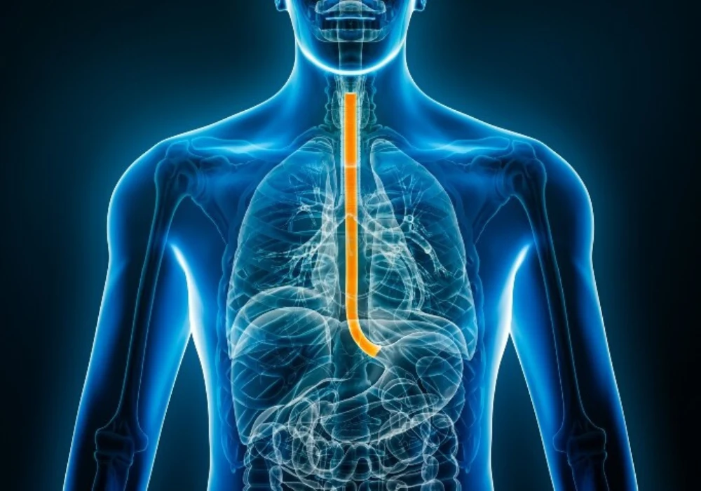
Anticipating postoperative outcomes in oesophageal squamous cell carcinoma (ESCC) remains difficult given frequent relapse and poor prognosis after recurrence. Contrast-enhanced CT contains quantitative information beyond routine visual assessment. By extracting radiomic features from preoperative CT and combining them with clinicopathological data, a machine learning framework can estimate 3-year recurrence risk and distinguish local-regional from distant relapse after radical surgery. A single-centre retrospective analysis developed such models and paired them with a nomogram for individual survival estimation, enabling risk stratification that was subsequently examined for associations with benefit from postoperative adjuvant therapy.
Cohort and Outcomes
A total of 526 patients underwent radical oesophagectomy with two-field lymph node dissection using Ivor-Lewis or McKeown procedures with comparable outcomes. Mean age was 63 years with 427 men and 99 women. Within three years, 125 patients relapsed: 14 local, 26 locoregional lymph node, 21 cervical lymph node and 64 distant metastases. Disease-free survival spanned surgery to recurrence, death or last follow-up without recurrence, and overall survival spanned surgery to death from any cause.
Baseline factors reflected established prognostic variables. Lymph node positivity, degree of differentiation, tumour length and neurovascular infiltration were evaluated for association with outcome. Stage distribution was 162 stage I, 193 stage II and 171 stage III. Neoadjuvant therapy was given to 54 patients and postoperative adjuvant therapy to 267 patients. All patients had preoperative cervical lymph node ultrasound and thoracic upper abdominal contrast-enhanced CT without clearly enlarged cervical nodes on imaging. The five-year survival rate across the cohort was 33.08%.
Model Development and Performance
Tumours were segmented on preoperative CT using 3D-Slicer. A total of 1702 radiomic features were extracted with PyRadiomics, reduced by t-test to 302, then refined via least absolute shrinkage and selection operator with 10-fold cross-validation using the 1-SE criterion. Six features were retained for recurrence prediction and nine for recurrence-pattern prediction. Clinicopathological variables were screened with Cox proportional hazards models, then integrated with radiomic features in Python to train logistic regression and support vector machines for 3-year recurrence and pattern classification. Class imbalance during training was addressed with synthetic minority over-sampling technique.
Must Read: AI and Hyperspectral Imaging for Early Oesophageal Cancer Detection
Patients were split 7:3 into training and validation cohorts (368 and 158). The combined models showed robust discrimination. In the training cohort, areas under the receiver operating characteristic curve (AUCs) were 0.826 for logistic regression and 0.820 for support vector machines. In the validation cohort, AUCs were 0.830 and 0.825. Compared with radiomics-only and clinicopathological-only approaches, the combined model performed better, with a net reclassification improvement of 0.127 and a confidence interval of 0.089–0.167.
Operating characteristics favoured reliable exclusion of recurrence. Sensitivity exceeded 91% in both cohorts and negative predictive value was greater than 91%. Specificity in the validation cohort reached 80.8% for logistic regression and 77.6% for support vector machines, while positive predictive values were modest at 46.2–50.0% consistent with class imbalance. Kaplan–Meier analyses showed longer disease-free and overall survival among patients predicted as non-recurrent. For recurrence-pattern classification, AUCs were 0.80 for logistic regression and 0.81 for support vector machines, supporting separation of local-regional from distant metastatic relapses.
A nomogram integrated the radiomics score with clinicopathological predictors to estimate 1-, 3- and 5-year survival. Calibration plots indicated close agreement between predicted and observed outcomes, and decision curve analysis supported clinical applicability. The workflow and codes for feature selection and nomogram calculation were provided in supplementary material.
Risk Stratification and Therapeutic Signals
Outputs from the classifier and nomogram were combined to define four risk classes, separating patients by recurrence probability and survival expectation. Propensity score matching within each class balanced clinicopathological covariates to compare outcomes between those receiving postoperative adjuvant therapy and those who did not. Survival differed significantly across the four classes, indicating effective stratification.
A clinically relevant subgroup emerged among stages II–III in the high-recurrence, low-survival class. In this group, postoperative chemotherapy was associated with improved disease-free survival with a hazard ratio of 0.372 and a 95% confidence interval of 0.206–0.669 with p < 0.001. No similar associations were identified in other strata after matching. High negative predictive value suggests an opportunity to reduce surveillance intensity for low-risk patients, while pattern prediction may guide selection of follow-up imaging aligned with likely sites of relapse.
Limitations included single-centre design, a primarily East Asian population and retrospective data that may introduce confounding. Detecting recurrence in early-stage disease may be more challenging as the proportion of T1–2N0 cases increases. Event counts limited more granular prediction of specific local sites such as anastomotic versus nodal recurrence. Future directions emphasised multi-centre validation, broader population testing and refinement of site-specific prediction.
Combining CT-derived radiomics with clinicopathological features enabled accurate prediction of 3-year recurrence and relapse pattern in ESCC after radical surgery, outperforming single-source models. The pairing of a high-performing classifier with a calibrated nomogram supported clinically meaningful risk stratification and identified a subgroup in which postoperative chemotherapy aligned with improved disease-free survival. These findings support structured postoperative planning that tailors surveillance and adjuvant therapy consideration to risk profiles derived from standard preoperative imaging and routine clinical data.
Source: Insights into Imaging
Image Credit: iStock


