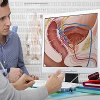
Combined hepatocellular cholangiocarcinoma (CHC) is a rare form of primary liver malignancy containing components of both hepatocellular carcinoma (HCC) and intrahepatic cholangiocarcinoma (ICC). Although the imaging features of CHC, HCC, and ICC on contrast-enhanced ultrasound (CEUS) may overlap, CEUS could be used in the differential diagnosis of CHC from HCC and ICC, according to new research published in the American Journal of Roentgenology.
"Marked washout in the late phase was a common feature of CHC and ICC that could be used to differentiate CHC and ICC from HCC," the study says. "Hyperenhancement in the arterial phase followed by marked washout in the late phase was the most common wash-in–washout pattern of CHC that could be used to distinguish CHC from ICC."
Investigators have reported that CHC shared many demographic and clinical features similar to ICC in Western case series, whereas other studies found that CHC showed intermediate clinical features with HCC and ICC. Because of these overlapping demographic and clinical features, as well as the indistinguishable clinical traits of CHC and poor prognosis of CHC, a correct diagnosis of CHC with imaging findings may have prognostic significance, particularly in determining transplant exception status and making management decisions.
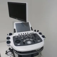
Meanwhile, CEUS has recently improved the diagnostic performance of ultrasound for several types of disease, especially in the differentiation of various types of hepatic lesions. The aim of the current study was to evaluate the diagnostic performance of CEUS in distinguishing CHC (type C) from HCC and ICC. This retrospective study included 33 patients with pathologically confirmed CHC and matched control subjects with pathologically confirmed HCC (n = 30) or ICC (n = 32) who underwent preoperative CEUS from January 2005 to December 2015. The CEUS images of the hepatic lesions were subjectively analysed in consensus by two radiologists. The diagnostic performances were evaluated by ROC analysis.
Researchers found that, in the arterial phase, hyperenhancement was more common in CHCs (76%) and HCCs (100%) than in ICCs (22%), whereas in the late phase marked washout was more common in CHCs (76%) and ICCs (100%) than in HCCs (10%). Using marked washout in the late phase to differentiate CHC from HCC, the area under the ROC curve (AUC) was 0.829, and the sensitivity, specificity, and accuracy were 78%, 90%, and 83%, respectively. Using hyperenhancement in the arterial phase followed by marked washout in the late phase to distinguish CHC from ICC, the AUC value was 0.663, and the sensitivity, specificity, and accuracy were 55%, 78%, and 66%.
"Using CEUS for the differentiation of CHC from ICC proved to be more difficult. A previous study with CT and MRI showed that the correct diagnosis of CHC was made in only a minority of cases. However, when the imaging features of both HCC and ICC are present in the same tumour after careful analysis of CT and MRI, the radiologist should recognise that CHC is a possible diagnosis," the researchers wrote.
The study has several limitations including that the range of lesion size was large and no comparison was made for the CEUS features between small and large lesions, "because of the rarity of CHC and the relatively small patient population," the research team note.
The researchers also said that further prospective studies are needed to compare the findings of CHC on CEUS, CT, and MRI.
Source: American Journal of Roentgenology
Image Credit: Max Powers
Latest Articles
Contrast-enhanced ultrasound, combined hepatocellular cholangiocarcinoma, intrahepatic cholangiocarcinoma
Combined hepatocellular cholangiocarcinoma (CHC) is a rare form of primary liver malignancy containing components of both hepatocellular carcinoma (HCC) and intrahepatic cholangiocarcinoma (ICC). Although the imaging features of CHC, HCC, and ICC on contr