A recent study of concussion patients using diffusion tensor imaging (DTI), entitled “Sex Differences in White Matter Abnormalities after Mild Traumatic Brain Injury: Localization and Correlation with Outcome”, found that females recovered faster after concussion than males did.
The findings are published online in the journal Radiology and show that DTI can be used as a bias-free way to predict concussion outcome.
In excess of 17 million Americans suffer from concussion each year, a mild traumatic brain injury (mTBI). Roughly 15 percent of these patients suffer persistent symptoms lasting longer than three months.
In order to assess concussion injury severity, outcome and recovery time physicians have typically been reliant on patient cooperation, and as Saeed Fakhran, M.D., assistant professor of neuroradiology at the University of Pittsburgh School of Medicine explained, often the MRI and CT brain images taken of concussion patients are normal. He praised diffusion tensor imaging as the first imaging technique capable of showing abnormalities associated with concussion, achievable due to its ability to visualise white matter tracts at a microscopic level.
The brain's white matter is composed of millions of nerve fibers called axons that act like communication cables connecting various regions of the brain. An advanced form of MRI that lets researchers assess microscopic changes in the brain’s white matter, DTI produces a measurement, called fractional anisotropy (FA), of the movement of water molecules along axons.
When white matter is healthy, this water movement is one-directional, rather uniform and high in FA measurement. More random water movement decreased the FA values, and abnormally low FA is linked to cognitive impairment in patients with brain injuries.
The study included imaging results and medical records of close to 70 patients diagnosed with mTBI between 2006 and 2013, almost two thirds were males with a median age of 17, whereas the female median age was 16. 68 percent of the male patients were injured while playing a sport, as were 45 percent of the females. The control group consisted of 21 people.
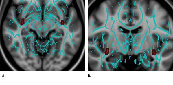
All study participants were subject to the same assessment, which included DTI of the brain and a computerised neurocognitive test. The DTI scans of the mTBI patients showed abnormalities within the uncinate fasciculi (UF), a white matter tract that connects the frontal and temporal lobes of the brain. Although its exact role is controversial, the UF tract is believed to allow temporal lobe-based memory associations to modify behavior though interactions with another area of the brain.
In comparison to the female mTBI patients, the males’ DTI scans had significantly decreased UF FA values. Dr. Fakhran stated that further research into the issue of gender and concussion would be required to determine who recovers faster and why.
Analysing the data statistically showed that UF FA value was a stronger predictor of recovery time than neurocognitive testing-based initial symptom severity, with decreased UF FA being the most substantial risk factor for a recovery time in excess of three months. Male gender also directly correlated with increased recovery time.
Dr. Fakhran explained the great clinical impact of the DTI and UF FA’s potential to predict outcome after concussion since MR scanners provide factual feedback. Patient reporting has so far been the main indicator, yet possibly driven by ulterior motives, such as wanting to get back to play, it has not always been reliable.
Regardless of initial symptom severity, all concussion patients had an average symptom recovery time of 54 days. Males reported a significantly longer mean time of 66.9 days, whereas females recovered in an average of 26.3 days.
Commenting on these results Dr. Fakhran stated that they indicated a potential role for UF FA values in triaging concussion patients in the future, and he added that there was prognostic value in DTI for both children participating in sports as well as for professional athletes.
He did caution that at this point DTI was not able to provide individual prognoses, however there was hope for this as lower FA values in the uncinate fasciculi could offer a metric for evaluating the severity of mild traumatic brain injuries and predict clinical outcome.
6 May 2014
Latest Articles
Research, Imaging, Concussion, gender, MR, DTI, mTBI, diffusion tensor imaging
A recent study of concussion patients using diffusion tensor imaging (DTI), entitled “Sex Differences in White Matter Abnormalities after Mild Traumatic...


