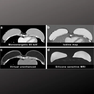
Breast reconstruction using silicone implants is a common cosmetic procedure for aesthetic enhancement, correcting congenital deformities, or restoring breasts after mastectomy in breast cancer patients. Over time, silicone implants can degrade due to gel cohesivity, surface structure, age, and location, leading to issues like microscopic silicone gel leakage or implant rupture. These complications can necessitate surgical removal of the implant or surrounding tissue and lymph nodes. Whole-body photon-counting CT (PCCT) is an emerging CT technology, but its utility for evaluating silicone breast implants in the thoracic region has not been studied extensively. A recent study published in European Radiology Experimental aimed to assess the contrast properties of silicone implants in thoracic PCCT and determine the best reconstruction technique for optimal image contrast. Additionally, the study evaluated the diagnostic accuracy of PCCT in detecting degenerative changes and ruptures in silicone breast implants, which is crucial for proper clinical management.
Detecting Implant Ruptures: The Limitations of Traditional Imaging Techniques
Intracapsular ruptures, occurring within the fibrotic capsule, are more common than extracapsular ruptures. They can be hard to detect clinically, via mammography, or ultrasound. Breast MRI with silicone-selective sequences is the most sensitive method for detecting implant ruptures, revealing signs like silicone collections, the linguine sign, or various other signs depending on the rupture type. Calcifications on the capsule can also form over time without necessarily indicating a rupture. Imaging techniques like computed tomography (CT) can also identify implant ruptures, but single-energy CT has limitations due to the similar radiodensity of silicone and breast tissue. Dual-energy CT and dedicated photon-counting breast CT offer better detection capabilities for silicone implant ruptures.
Study Overview: Cohort and Implant Characteristics
A prospective cohort study was conducted at the University Medical Center Freiburg, Germany, from January 2022 to May 2023. The study included 21 female patients with a total of 29 silicone breast implants who required thoracic CT imaging and had available breast MRI studies. The patients' ages ranged from 26 to 81 years at the time of implantation (mean age: 47 years) and from 41 to 85 years at the time of the study (mean age: 60 years). The implants had been in place for 2 to 24 years (mean duration: 10 years). The reasons for implantation varied, including breast removal after ipsilateral or bilateral breast cancer, protective removal with reconstruction after contralateral breast cancer, and breast augmentation. Most of these implants were textured or polyurethane foam-filled prostheses.
Contrast Properties and Diagnostic Accuracy of Thoracic PCCT
Silicone breast implants displayed clear contrast properties in photon-counting computed tomography (PCCT), with high contrast-to-noise ratios (CNRs) for both implant-to-muscle and implant-to-fat in the iodine map. Virtual unenhanced and monoenergetic reconstructions also showed high CNRs for specific contrasts. PCCT proved highly accurate in identifying degenerative changes and ruptures in silicone breast implants. It had very high diagnostic accuracy for detecting signs like the linguine sign, intraimplant fluid, peri-implant silicone collections, and high diagnostic accuracy for the keyhole sign and membrane folds.
Challenges in Breast Cancer Screening and Alternative Assessment Methods
Silicone breast implants often pose challenges in breast cancer screening and can lead to complications like capsular contracture or rupture. While mammography, tomosynthesis, and ultrasound have limitations, MRI offers high sensitivity but is costly, time-consuming, and not universally available. Alternative methods for assessing silicone breast implants include dual-energy CT and dedicated breast CT. PCCT, an emerging technology, offers advantages like higher spatial resolution, improved iodine signal, artifact reduction, and multienergy imaging with reduced radiation dose.
Thoracic PCCT Imaging: Accuracy and Potential Applications
The study found that thoracic PCCT imaging, especially in the prone position, accurately identifies signs of degenerative changes and ruptures in silicone breast implants. It can detect collapsed and uncollapsed implant ruptures, though some signs like folds of the membrane and peri-implant fluid collections had lower diagnostic accuracy possibly due to imaging differences compared to MRI. PCCT also enables three-dimensional visualization of breast tissue with contrast agent administration, aiding in identifying or excluding suspicious lesions. It could potentially be used for whole breast evaluation in the future, including lymph node assessment.
In conclusion, despite PCCT devices not being widely available yet, this pilot study demonstrated that thoracic PCCT offers promising results in assessing silicone breast implants and diagnosing implant integrity, eliminating the need for dedicated breast CT systems or more expensive and time-consuming breast MRI.
Source & Image Credit: European Radiology Experimental


