HealthManagement, Volume 25 - Issue 3, 2025
Moscow researchers have developed advanced medical phantoms to enhance ultrasound and MRI training, including liver, prostate, foetal and spinal models. Backed by the MosMedMaterial materials database, these phantoms improve diagnostic accuracy and procedural skills. International collaborations support AI development and protocol optimisation. The initiative strengthens medical education, clinical research and digital healthcare innovation.
Key Points
- Moscow developed realistic phantoms to improve ultrasound and MRI diagnostic training.
- The liver phantom helps distinguish benign from malignant lesions and guide procedures.
- MosMedMaterial offers materials data for building accurate, multimodal imaging phantoms.
- International collaboration supports AI algorithm development using these phantoms.
- The initiative enhances clinical skills, protocol design and scientific self-sufficiency.
Phantom-Based Training for Ultrasound Imaging
Researchers in Moscow have created a liver phantom encased in soft tissue to assist in training physicians in ultrasound. The phantom includes reconstructed blood vessels and 18 inclusions representing cysts, malignant tumours and adenomas. This setup generates realistic ultrasound images, helping doctors improve their ability to identify liver diseases and perform medical procedures.
Anastasia Rakova, Deputy Mayor of Moscow for Social Development, reported on this advancement, emphasising that training on phantoms is a vital factor in significantly improving the quality of medical care in Moscow. She noted that doctors who practice on test-objects until their skills become automatic demonstrate greater confidence and precision when performing procedures on patients. Russian-developed ultrasound phantoms now provide images closely resembling real organs, thereby enhancing the effectiveness of medical training. According to Rakova’s report, scientists in Moscow have created 12 phantoms. The latest liver phantom allows practitioners to distinguish between benign and malignant liver lesions and perform ultrasound-guided interventions.
Development of Phantom Models for MRI Applications
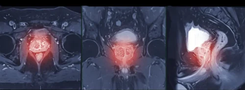
Each of the models has unique properties. The prostate phantom improves magnetic resonance imaging (MRI) accessibility, particularly for patients with metal-on-metal hip implant systems. Previously, scanner configuration required patient participation, but the phantom now enables pre-scan setup, reducing errors and streamlining the process. Additionally, developing new scanning protocols for patients with implants was time-consuming, prolonging procedure times and disrupting clinic schedules. The phantom mitigates these issues by allowing advance calibration, enhancing study accuracy.
Another model, the foetal phantom, optimises imaging protocols for foetal MRI. MRI provides high-resolution, high-contrast imaging of the foetus, abdomen and pelvis without ionising radiation, making it a valuable adjunct to ultrasound for detecting various pathologies. However, foetal MRI protocol is challenging. Given its critical role in pregnancy management and planning of intrauterine interventions, optimising imaging protocols is essential. The foetal phantom facilitates this process, enhancing diagnostic precision and clinical decision-making.
To optimise MRI scanning protocols, clinicians often rely on pregnant volunteers. Although safe for both mother and foetus, prolonged scanning in uncomfortable positions is required due to foetal movement and the mother's breath, which degrade image quality and reduce diagnostic value. To address these challenges, a foetal phantom was developed in collaboration with the Kulakov National Medical Research Center for Obstetrics, Gynaecology and Perinatology, enabling protocol refinement without patient burden.
The lumbosacral phantom is an anatomically accurate simulation model designed to enhance ultrasound-guided needle insertion skills for anaesthesiologists, particularly in chronic pain management. Derived from anonymised human CT data, the phantom replicates two vertebrae, the coccyx, iliac bones and surrounding soft tissues using durable materials. Its inclusion of the sacrum enables training for specialised procedures, such as caudal anaesthesia. Additionally, this phantom aids preoperative planning and, when combined with augmented reality (AR), improves visualisation of critical structures (eg blood vessels, nerves and tumours), refining procedural accuracy.
International Collaborations and AI Integration
Phantoms developed by Moscow researchers represent advanced simulation models that have garnered significant international interest. Notably, a breast phantom designed to enhance ultrasound-guided diagnostic and biopsy training for breast neoplasms has demonstrated its utility in a collaborative study between Russian and Chinese scientists. This research focuses on improving early breast cancer detection.
In April 2025, the Center for Diagnostics and Telemedicine and Beijing University of Technology (BJUT) initiated a strategic partnership to develop AI-based algorithms aimed at optimising ultrasound image quality and diagnostic precision, further validating the clinical relevance of these phantom models.

MosMedMaterial: Russia’s Phantom Material Database
The development of medical imaging phantoms has been ongoing for seven years at the Laboratory of the Center for Diagnostics and Telemedicine of the Moscow Healthcare Department. The Center has established Russia’s first database – MosMedMaterial – containing information on 23 solid and 19 liquid materials suitable for phantom creation. This database is regularly updated as new materials and data become available, supporting the advancement of medical training simulators.
Yuri Vasilev, Chief Consultant for Radiology of the Moscow Healthcare Department, reported the MosMedMaterial database as a valuable practical resource for clinicians and researchers. It consolidates information on materials appropriate for creating medical phantoms, enabling even small research teams to independently produce realistic organ models for their studies. Vasilev highlighted this as a significant step toward advancing medical education and research.
The developers systematically evaluated each material included in the database for its capacity to mimic specific organs or tissues across various imaging modalities, including computed tomography, magnetic resonance imaging and ultrasound. For solid materials, mechanical properties were also thoroughly characterised. To enhance user accessibility, software with a search function based on organ type or material parameters has been developed. Additionally, an interactive atlas is currently under development, systematising data on materials and their biological counterparts, with visual search capabilities using a three-dimensional human model. This atlas will allow users to visually explore a three-dimensional model of the human body, streamlining the identification and selection of suitable materials for biomedical and imaging purposes.

Anton Vladzymirskyy, Dr. Sc. in Medicine, MD, Deputy Director for Research at the Center for Diagnostics and Telemedicine, noted that most phantom developers traditionally rely on experimental researches to select materials, a process that is both time-consuming and resource-intensive. The Center’s systematic evaluation ensures that materials not only match the imaging characteristics of biological tissues but also replicate their mechanical properties, which is critical for effective medical training.
The established database enables a more technological approach to phantom development. Researchers in Moscow are optimising this process by utilising substances that replicate various human tissue types, allowing rapid modelling of organs and systems through combinations of existing materials.
The Center’s approach also revealed that some materials exhibit multimodal properties, suggesting promising potential for developing phantoms suitable for complex, multimodal diagnostic training.
Multimodal Phantom and Research Integration
The Center has developed the first multimodal arterial vessel phantom. This innovation forms part of a comprehensive pulsation simulation system designed for vascular imaging studies using both computed tomography and ultrasound. This innovation supports the refinement of imaging protocols, serves as a simulation tool for medical education and facilitates research on blood flow modeling in diagnostic imaging. The technology accurately replicates vascular biomechanical, hydrodynamic and visual characteristics, enabling simulation of diverse vessel states.
Data from the MosMedMaterial database has already facilitated the rapid development of a forearm tissue model for scientific research conducted at the Department of Medical and Technical Information Technologies, Bauman Moscow State Technical University. Specifically, a model with predefined dimensions was required to validate the accuracy of a novel ultrasound-based method for assessing muscle function. The use of human volunteers was not feasible, as precise measurements of tissue dimensions in vivo are unattainable, thereby precluding rigorous method validation. In the absence of such control data, the reliability of the verification process would be compromised. The successful use of this phantom was reported in a scientific article published by MDPI (Kapravchuk et al. 2025).
Olga Omelyanskaya, CAO of the Research and Development Department at the Center for Diagnostics and Telemedicine, remarked that the creation of a soft tissue phantom of the forearm exemplified effective collaboration between two leading scientific teams in Moscow. She emphasised that the partnership between the Center for Diagnostics and Telemedicine and Bauman Moscow State Technical University not only highlighted the development of advanced technologies by Moscow-based specialists but also demonstrated their capability to independently design and implement robust testing tools utilising an in-house database of materials for radiology diagnostics. Omelyanskaya further noted that such comprehensive, full-cycle projects form the foundation of scientific and technological independence.
Scientific Leadership and Output
The Center for Diagnostics and Telemedicine is a leading scientific and practical institution within the Moscow Healthcare Department. It oversees the management of radiology departments, drives digital healthcare transformation, implements AI technologies in clinical practice, conducts scientific research and trains medical professionals. Since 2013, the Center’s staff have produced over 800 scientific publications—including articles, methodological guidelines, monographs and training manuals,—registered more than 200 intellectual property results and developed 12 medical phantoms.
Conflict of Interest
None.
References:
Kapravchuk V, Ishkildin A, Briko A et al. (2025) Method of Forearm Muscles 3D Modeling Using Robotic Ultrasound Scanning. Sensors, 25(7):2298.


