One of the main organs affected by the coronavirus is the lung. While most patients have no symptoms or mild symptoms, some develop dyspnoea, tachypnea, and respiratory distress that may require mechanical ventilation.
In many parts of Europe, the US, and Asia, the CT scan has been widely used to assess the lungs in patients with COVID 19. Not only is the CT scan very specific but it can delineate the very fine features of the disease and can be used to follow up patients. However, most radiological societies discourage the use of CT scan as a screening tool because not only is it expensive but also time-consuming. They recommend the use of the CT scan only in patients with severe COVID-19 or those with ARDs.
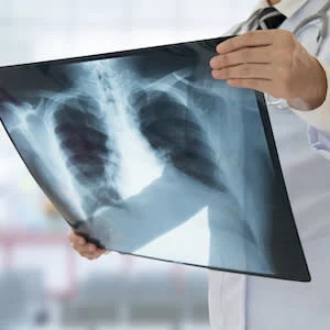
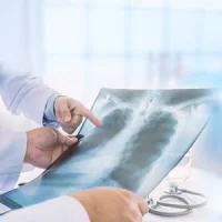
Chest radiograph (CXR) on the other hand, is widely available, and is cost-effective and portable. However, not much has been written about this technique and whether any patterns of disease exist. CXR has become one of the most useful tools in the evaluation of patients who develop COVID 19. While many studies have reported CXR changes in COVID 19 patients, no large scale study has been conducted to determine if there are any pathognomonic patterns on the chest radiograph.
In this retrospective study, researchers study the chest radiograph of COVID-19 patients to determine if it could aid in the clinical diagnosis of the infection. The study was undertaken in New Orleans in the middle of March 2020, during which time there was an exponential outbreak of the infection. CXRs of 366 COVID-19 patients were reviewed together with their RT-PCR tests. Each CXR was evaluated by two experienced radiologists for characteristics, and negative or nonspecific appearance of COVID-19. The COVID-19 imaging patterns were then compared against RT PCR results to determine if the chest radiograph could be used for accurate diagnosis.
In the study, there were 178 males (49%) and 188 females (51%) with a mean age of 52.7 years. About 10% (37) CXR exams revealed the characteristic COVID-19 appearance, 215 (57%) exhibited non-specific appearance, and 124 (33%) were considered negative for any lung abnormality. Of the 376 RT PCR tests done, 200 (53%) were positive and 176 were negative. The RT PCR tests took an average of 2.5 days for a result. The sensitivity and specificity for correctly identifying COVID 19 with characteristic CXR patients were 15.5% (31) and 96.6% (170), with PPV and NPR 83.8% and 50.1, respectively.
The conclusion of the study was that the presence of patchy and/or confluent band like ground-glass opacity or consolidation in the mid to lower lung and peripheral zone distribution on the CXR was highly suggestive of SARS-CoV2 infection and could be used in conjunction with clinical reasoning to make a diagnosis.
It is important to be aware that the chest radiograph as very low sensitivity for COVID 19 and should not be a substitute for the RT PCR test. The study has several limitations including no mention of other comorbidities in these patients which could have also presented with abnormalities on the CXR. Plus the image quality and technique were not controlled for and the broad category of the nonspecific category may have missed many cases of COVID 19, which may have been identified by CT scan.
Finally, a bilateral patchy and confluent band like ground-glass opacity or consolidation is not specific for COVID-19 and has a big differential diagnosis. CXR should only be used to make a tentative diagnosis of COVID-19 when serologic testing is not available.
Source: Radiology: Cardiothoracic Imaging
Image Credit: iStock
References:
Smith et al. (2020) A Characteristic Chest Radiographic Pattern in the Setting of COVID-19 Pandemic. Radiology: Cardiothoracic Imaging, 2(5).

