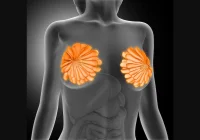
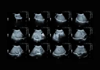
Breast ultrasound (US) is an indispensable diagnostic tool for assessing breast abnormalities. It has progressed significantly over the years, now complemented by advancements such as Shear Wave Elastography (SWE) and texture analysis. However, there are questions about the added value of these newer techniques, particularly in their ability to improve the differentiation between benign and malignant breast masses. Recent studies have explored the efficacy of ultrasound, SWE and texture analysis in breast cancer diagnostics.
Grayscale Ultrasound and BI-RADS Classifications
Grayscale ultrasound has long been the cornerstone of breast imaging, aiding in detecting and evaluating suspicious masses. The American College of Radiology developed the Breast Imaging Reporting and Data System (BI-RADS) to standardise breast imaging terminology, including categories ranging from benign (BI-RADS 1 and 2) to highly suggestive of malignancy (BI-RADS 5). Grayscale features such as mass shape, margin, and calcifications are key descriptors used to guide clinical decision-making. Masses within BI-RADS 4, for instance, may appear suspicious but do not have definitive signs of malignancy, often leading to biopsies.
Despite its effectiveness, grayscale ultrasound has limitations in specificity. It sometimes lacks the precision to differentiate subtle variations between benign and malignant masses, making complementary technologies like SWE appealing.
Shear Wave Elastography: Enhancing Specificity
Shear Wave Elastography (SWE) enhances the diagnostic power of traditional ultrasound by measuring tissue stiffness. Malignant tissues tend to be stiffer than benign ones, and SWE provides a quantitative assessment of this stiffness. Studies indicate that SWE can improve diagnostic specificity, particularly when combined with grayscale ultrasound. For example, stiffness values greater than 40 kPa on SWE often indicate malignancy, providing additional information beyond morphology alone.
In clinical practice, SWE is a valuable adjunct to conventional ultrasound. It allows radiologists to detect malignancies that may not present typical characteristics on grayscale ultrasound. However, as with any technology, its utility depends on the skill of the operator and the interpreting radiologist.
Texture Analysis: Does it Add Diagnostic Value?
Texture analysis, a subset of radiomics, offers a way to quantify subtle textural patterns within a breast mass that may not be visually apparent. By transforming imaging data into a series of quantifiable metrics such as voxel intensity, texture analysis could potentially enhance the diagnostic capability of breast ultrasound.
However, recent findings suggest that texture analysis may not significantly improve the differentiation of benign and malignant masses compared to traditional ultrasound features. In the case of breast cancer, the inclusion of texture analysis did not provide additional predictive value in differentiating malignancy from benign conditions. Other factors, such as the shape of the mass and the stiffness detected by SWE, remained the most reliable indicators of malignancy.
While texture analysis shows promise in other areas of radiology, its role in breast cancer diagnosis remains limited. Grayscale ultrasound, paired with Shear Wave Elastography, continues to be the most effective approach for distinguishing between benign and malignant breast masses. SWE, in particular, has proven to be a critical tool in enhancing diagnostic specificity. Future advancements in imaging technology may refine these methods further, but for now, traditional ultrasound features and elastography remain the gold standard in breast cancer screening.
Source: Journal of Breast Imaging
Image Credit: iStock


