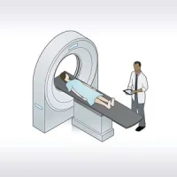
Researchers compared results from twenty patients with SAH, who underwent a conventional brain MRI and a SyMRI on a 3T MRI machine.
You might also like this: As more clinicians and patients are requesting a radiation-free technique to deal with cancer screening and management, whole-body magnetic resonance imaging (WB MRI) offers such a solution. This study reviewed its technical basis, current international guidelines for use and key applications.Learn more.
Key Findings
- SyMRI detected intracranial complications of SAH similarly to conventional MRI.
- SyMRI acquisitions have quality metrics comparable to conventional MRI.
- SyMRI acquisition time is shorter compared to conventional MRI.
The two techniques performed well in detecting ischaemic lesions and extra-axial collections (kappa = 0.80 and 0.88 respectively) and were good for the detection of hydrocephalus (kappa = 0.69). No significant differences were detected in the number of ischaemic lesions (p=0.31) or in the Evans index (p=0.11).
The WMv and CSFv measures were also similar (p=0.18 and p=0.94, respectively), as well as the volume of ischaemic lesions (p=0.79). The SyMRI acquisition time was shorter compared to conventional MRI regardless of the number of sections (32% and 6% time reduction for 4 or 3 mm section thickness, respectively).
Study results were published in Clinical Radiology June 27, 2021.
Source: Clinical Radiology
Photo: iStock
References:
Montejo C, Laredo C, Llull L, et al. (2021) Synthetic MRI in subarachnoid haemorrhage. Clinical Radiology. Published: June 27, 2021. DOI: https://doi.org/10.1016/j.crad.2021.05.021

