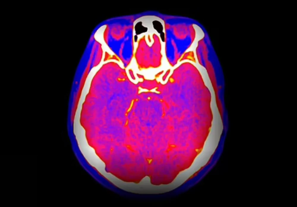
Spectral computed tomography (CT) is increasingly available across healthcare systems, offering energy-resolved information that can refine tissue characterisation and support quantitative assessment. Despite this growing access, consistent day-to-day use remains limited and quality assurance (QA) practices vary widely. A global survey coordinated by the International Atomic Energy Agency documented current availability, utilisation, QA and training patterns, highlighting progress alongside persistent barriers. The responses, gathered between October 2024 and June 2025 from institutions across multiple regions, indicate rapid modernisation of CT fleets, expanding clinical applications and uneven integration into routine workflows. Training gaps, especially for medical physicists and heterogeneous QA approaches were recurrent themes, pointing to the need for clearer guidance, aligned protocols and targeted capacity building to translate capability into consistent practice.
Access Expands but Adoption Remains Uneven
Respondents spanned Asia-Pacific, Europe, Latin America, Africa and the United States, with the largest participation from Asia-Pacific and Europe. Interest from countries with traditionally limited imaging infrastructure signalled widening awareness of spectral capability beyond well-resourced centres. Reported installations ranged from 2009 to 2025, with nearly half placed in the last four years, underscoring active fleet renewal. Across 168 spectral-capable systems, a subset were photon-counting CT scanners, most deployed from 2024 onwards. Systems from several vendors were represented, with one manufacturer accounting for the majority share and others covering the remainder. Facility-level capability varied markedly, from sites with no spectral-capable scanners to those operating fully spectral fleets, illustrating heterogeneous access despite overall diffusion.
Routine use lagged behind availability. Many scanners applied spectral protocols in only a small proportion of examinations. A sizeable segment reported use in fewer than 1% of weekly scans, and another share in fewer than 10%. About a third indicated use across 10–50% of examinations, while only a small minority exceeded 50%. The need to select spectral modes prospectively on certain systems was cited as a practical constraint in high-throughput environments, where workflow efficiency and scan time are closely managed. These patterns suggest that access alone does not guarantee integration and that operational demands often determine whether spectral options are activated beyond targeted indications.
Training and QA Gaps Constrain Consistency
Training emerged as a central determinant of use. Radiologists had received training in a number of institutions, with a similar number reporting none and a portion not responding. For medical radiation technologists, training coverage was broader yet still incomplete. The most pronounced shortfall concerned medical physicists, with fewer institutions reporting trained physicists than those reporting none. Given spectral CT’s technical demands, including protocol design, reconstruction choices, post-processing and dose considerations, the imbalance highlights a priority area for workforce development.
Must Read: Spectral CT Reveals Airway Differences in Pneumonia Subtypes
QA and quality control (QC) practices lagged technology uptake. More than half of respondents reported the absence of spectral-specific QC tests. Only a smaller share indicated that such tests were in place, with the remainder not responding. Dosimetry approaches often remained unchanged when moving from conventional to spectral modes, with only a minority reporting different dose assessment methods. Where QC was performed, frequency and method varied from daily or weekly calibrations to periodic constancy testing using vendor or third-party phantoms. Some sites relied on automated consistency checks, while others followed general CT routines without spectral-specific procedures. Existing guidance was perceived as outdated for modern systems, and awareness of available multi-energy CT QC frameworks was uneven. Without harmonised QA/QC standards and aligned training, institutions risk inconsistent image quality, suboptimal protocol selection and limited confidence in quantitative outputs.
Clinical Uses Grow Amid Workflow Friction
Reported applications were diverse, spanning cardiology, abdominal and pancreatic imaging, urology, musculoskeletal assessment, neurological indications and oncology. Common use cases included pulmonary embolism assessment, particularly where contrast load must be minimised, abdominal lesion characterisation, perfusion evaluation, low-contrast-dose scenarios and kidney stone analysis with differentiation of uric acid and calcium composition. Cardiac planning and other cardiac imaging tasks featured in several responses. Functional tools such as virtual non-contrast imaging, metal artefact reduction, iodine mapping and interventional planning were also noted. Some centres applied spectral modes broadly across most protocols, while others reserved them for narrow indications or reduced use due to operational constraints.
Free-text responses outlined why broader deployment remains challenging. Knowledge gaps and uncertainty about interpreting spectral outputs limited clinician confidence. Perceived clinical relevance varied by setting, with limited demand in services focused on streamlined pathways. Technical and workflow friction was common, including lack of post-processing software, limited compatibility with newer tools, the need for separate acquisition modes on specific scanners and additional review time. Concerns about radiation dose were mentioned infrequently, suggesting dose was not a dominant barrier in the sample. Administrative and operational factors also appeared, with examples of software features being locked or considered of low added value. Funding or reimbursement issues were not explicitly identified among the reported barriers.
Altogether, the findings indicate that the value of spectral CT depends on more than hardware. Successful adoption requires clear protocols, accessible post-processing, interoperable tools and staff confidence in the clinical utility of outputs. High patient volumes amplify these needs, as even modest increases in reconstruction or interpretation time can disrupt schedules and reduce throughput.
Global access to spectral CT is expanding, including in regions with historically limited imaging resources, yet routine use trails due to training shortfalls, variable QA/QC practice and workflow friction. Clinical applications are broad and growing, but consistent integration depends on practical enablers that reduce operational burden and standardise quality. Targeted training, particularly for medical physicists, together with updated, harmonised guidance for QA/QC and dose assessment, can help ensure that expanding capability translates into reliable practice. Coordinated efforts by professional bodies, manufacturers and healthcare institutions will be essential to embed spectral CT into everyday pathways in a way that supports diagnostic quality, operational efficiency and patient care.
Source: Insights into Imaging
Image Credit: iStock


