According to a new
study being presented at the annual meeting of the Radiological Society of
North America (RSNA), digital breast tomosynthesis, also known as 3D
mammography, has the potential to significantly increase the cancer detection
rate in mammography screening of women with dense breasts.
Breasts that have more fibrous or glandular tissue but not much fatty tissue are considered to be dense. Research indicates that dense breasts are more likely to develop cancer. However, cancer in dense breasts can be difficult to detect on mammograms.
Imaging modalities like ultrasound and MRI are often used to help find cancers that cannot be seen on mammograms, but both modalities have higher rates of false-positive findings, resulting in additional tests and unnecessary biopsies. This results in making MRI and ultrasound expensive to implement in high-volume screening programs, according to the study's lead author Per Skaane, MD, PhD, from the Department of Radiology at Oslo University Hospital in Oslo, Norway.
Dr. Skaane and colleagues evaluated tomosynthesis as a promising breast cancer screening option that has the potential to address some of the limitations of mammography by providing 3D views of the breast.
"Tomosynthesis could be regarded as an improvement of mammography and would be much easier than MRI or ultrasound to implement in organized screening programs," Dr. Skaane said. "So the intention of our study was to see if tomosynthesis really would significantly increase the cancer detection rate in a population-based mammography screening program."
During the study, the research team compared cancer detection using full-field digital mammography (FFDM) versus FFDM plus digital breast tomosynthesis. The study participants included 25,547 women between the ages of 50 and 69. Breast density was classified based on the American College of Radiology's Breast Imaging-Reporting and Data System (BI-RADS), with 1 being the least dense and 4 being the most dense.
In the study group, 257 malignancies were detected using FFDM or a combination of FFDM and tomosynthesis. This included 105 in the density 2 group and 110 in the density 3 group. 82 percent of the cancers were detected with FFDM plus tomosynthesis, while 63 percent were detected with FFDM alone. FFDM plus tomosynthesis pinpointed 80 percent of the 132 cancer cases in women with dense breasts, compared to only 59 percent for FFDM alone.
"Our findings are extremely promising, showing an overall relative increase in the cancer detection rate of about 30 percent," Dr. Skaane said. "Stratifying the results on invasive cancers only, the relative increase in cancer detection was about 40 percent."
Overall, the study showed that tomosynthesis not only improved the cancer detection rate in women with dense breasts, it also helped increase detection for women in the "fatty breast" BI-RADS categories, improving the cancer detection rate from 68 percent to 84 percent.
"Our results show
that implementation of tomosynthesis might indicate a new era in breast cancer
screening," Dr. Skaane said.
Skaane observed that interpreting a tomosynthesis image takes around 80-90 seconds, whereas for 2D images it's around 25 seconds per woman. However, tomosynthesis picks up more cancers.
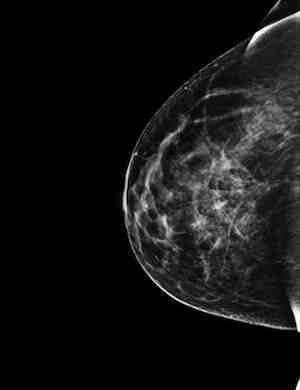
Figure 1. 46-yr-old woman 2D full-field digital mammography view of breast.
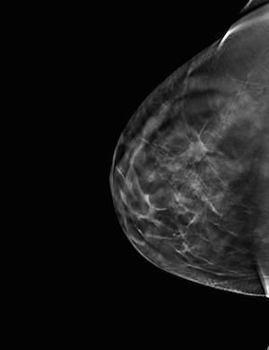
Figure 2. 46-yr-old woman 3D digital tomosynthesis view of breast.
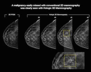
Figure 3. A
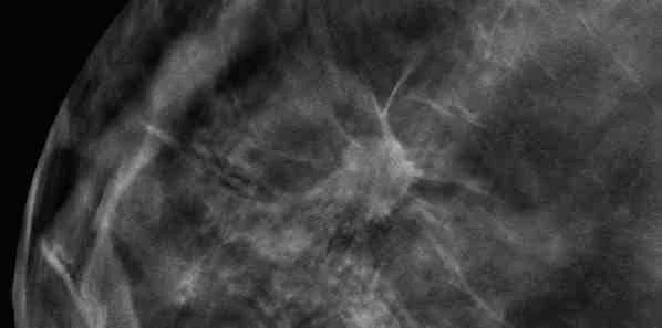
46-year-old woman’s routine screening mammogram: 2D mammogram is
essentially negative. 3D Tomosynthesis images reveal a 15 mm spiculated
mass between slices 38-48, diagnosed as invasive ductal carcinoma,
grade 2.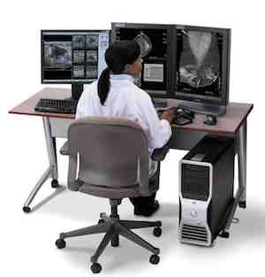
Figure 4. A radiologist reading a 3D mammogram on a diagnostic workstation.
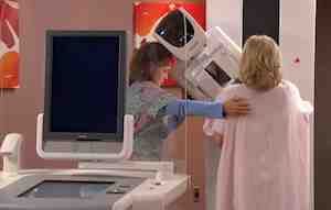
Figure 5. A patient being imaged with the 3-D mammography system.
Figure 6. Sample digital tomosynthesis image.
Video
Videos, including discussions and patient education, are available on the RSNA press page.
Caption on main image: Spiculated mass on 3-D tomosynthesis image.
Source: RSNA
Image Credit: RSNA

