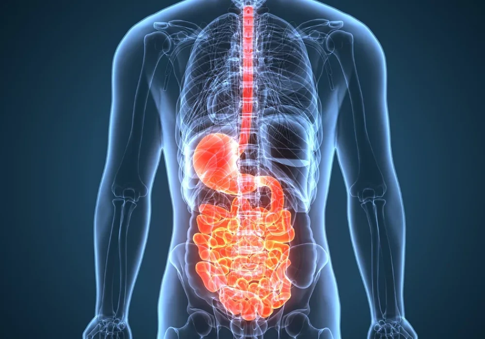
Crohn’s disease is a chronic inflammatory condition with remitting and relapsing activity that often requires repeated imaging to assess extent, complications and treatment response. Endoscopy and biopsy underpin diagnosis, yet cross-sectional imaging with ultrasound, computed tomography and magnetic resonance is pivotal for mapping disease and guiding care. Because many patients undergo multiple examinations across a lifetime, cumulative exposure to ionising radiation can become substantial, particularly when computed tomography is used during acute episodes. Balancing diagnostic yield with radiation safety has therefore become central to imaging pathways. Recent advances in ultrasound and magnetic resonance, together with dose-optimised computed tomography techniques, offer practical routes to reduce exposure while maintaining diagnostic quality.
Imaging Roles and Trade-offs in Crohn’s Care
A range of modalities supports diagnosis and surveillance, including plain radiography, fluoroscopy, ultrasound (US), computed tomography (CT), magnetic resonance imaging (MRI) and nuclear medicine techniques. Traditional fluoroscopic studies have declined with the rise of cross-sectional imaging but still offer dynamic assessment, albeit with limitations in detecting extraluminal disease and longer, less comfortable procedures. US is widely available, non-invasive and free of ionising radiation. It can assess bowel wall thickness, vascularity and mesenteric changes and with oral contrast or contrast-enhanced ultrasound can help distinguish active from quiescent inflammation. Intestinal ultrasound is increasingly integrated as a point-of-care assessment, though performance remains operator-dependent and sustained training is required.
CT is well established for detecting disease extent and complications such as abscesses, fistulae and strictures, and CT enterography enhances small-bowel evaluation with large-volume oral contrast and high-quality multiplanar reconstructions. CT also guides percutaneous drainage, potentially avoiding surgery in a proportion of patients. MR enterography provides comparable assessment of inflammatory activity without ionising radiation and can incorporate dynamic sequences to evaluate motility and characterise strictures, although longer acquisition times, motion artefact and availability constraints are recognised. In acute presentations, CT often remains the first choice due to speed, tolerance and whole-abdomen coverage. Positron emission tomography with CT or MRI has been explored for inflammatory activity, but high cost, availability issues and, for PET/CT, added radiation limit routine use.
Must Read: Managing Radiation Exposure in ICU Imaging
Cumulative Dose and Drivers of Exposure
Ionising radiation can damage DNA through free radical formation, and exposure is commonly expressed as effective dose in millisieverts. A CT of the abdomen and pelvis is quoted at approximately 8 mSv, about twice normal annual background radiation. Because Crohn’s disease often requires repeat cross-sectional imaging to evaluate flares, complications and response to therapy, lifetime cumulative effective dose can be high. Reports indicate that roughly 15–25% of patients may accrue 50–75 mSv over their disease course. Across cohorts, CT is consistently identified as the dominant source of exposure, contributing around three-quarters of cumulative dose.
Higher exposure correlates with more severe or extensive disease, complications and surgical treatment. Patients receiving immunosuppressive therapy can also undergo more follow-up imaging to assess response. Body composition influences dose during abdominopelvic CT, with adipose tissue volume a strong predictor of exposure. Access to MRI further shapes practice. Availability varies widely between countries, and limited provision can shift imaging towards CT, increasing cumulative exposure for local populations. These factors underscore the need for careful modality selection and dose-aware pathways, particularly in younger patients who are more sensitive to radiation.
Practical Ways to Minimise Dose
Guideline recommendations support MR enterography and ultrasound over CT for follow-up where local expertise permits, given comparable diagnostic performance and absence of ionising radiation. Imaging frequency should be critically reviewed to avoid unnecessary examinations, applying ALARA principles. For CT referrals, senior clinical oversight can help ensure that scans are justified, especially during acute exacerbations where utilisation has risen without a corresponding increase in clinically significant findings.
Dose-tracking software can assist by surfacing a patient’s cumulative exposure across radiography, fluoroscopy, CT and nuclear medicine, informing modality choice at the point of care. When CT is indicated, modern dose-optimisation strategies—limited scan range, protocol tailoring to the clinical question, automatic tube current modulation and judicious kV selection—can maintain diagnostic quality at lower dose. Iterative reconstruction has enabled substantial reductions compared with filtered back projection, and newer AI-based image enhancement further suppresses noise on low-dose studies. Photon-counting CT is an emerging technology that measures individual photon energies, improving spatial and contrast-to-noise performance at lower dose and showing early promise in characterising inflammatory severity, although cost and availability currently constrain adoption.
A case-by-case approach remains essential. In suspected Crohn’s disease, MR enterography and intestinal ultrasound are preferred for comprehensive characterisation, where accessible. In emergency presentations, CT or CT enterography is often necessary for rapid, whole-abdomen evaluation and to exclude alternative diagnoses. During stabilised follow-up, ultrasound can monitor for activity, reserving MR enterography for more detailed assessment or equivocal findings. Across settings, radiologists can support referrers with modality selection and dose-optimised protocols aligned to local resources.
Patients with Crohn’s disease face heightened risk of high lifetime radiation exposure due to repeat imaging, with CT the main contributor. Diagnostic certainty and timely intervention remain paramount, yet pathways can meaningfully reduce dose by prioritising ultrasound and MR enterography for surveillance, scrutinising imaging frequency and deploying modern CT optimisation when CT is required. Dose tracking, iterative and AI-enabled reconstruction, and emerging photon-counting CT offer practical and future-facing tools to sustain image quality at lower exposure. Embedding these measures into routine practice aligns imaging strategy with long-term patient safety without compromising clinical decision-making.
Source: British Journal of Radiology
Image Credit: iStock


