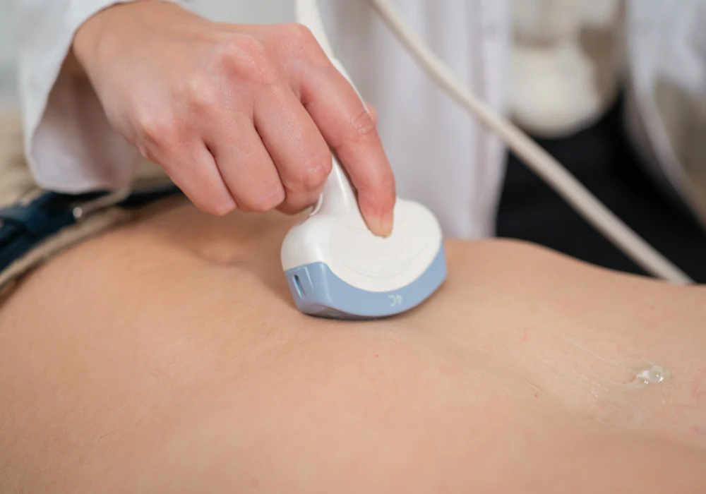
Solid-cystic liver lesions comprise a wide spectrum of pathological entities, including benign conditions, borderline tumours and malignant neoplasms. Accurate characterisation of these lesions is essential for guiding clinical management, as the treatment and prognosis vary considerably depending on the underlying diagnosis. While conventional ultrasound (US) is often the first imaging modality employed, its diagnostic limitations are well recognised. Contrast-enhanced ultrasound (CEUS) has emerged as a promising alternative, offering real-time assessment of vascularity and tissue perfusion. A recent study aimed to assess the performance of CEUS in comparison with conventional US and contrast-enhanced computed tomography (CECT), analysing 157 liver solid-cystic lesions of various origins.
Diagnostic Complexity of Solid-Cystic Liver Lesions
Liver solid-cystic lesions encompass a broad array of conditions ranging from simple cysts and abscesses to complex parasitic infections, borderline neoplasms such as intraductal papillary neoplasms of the bile duct (IPNB), and malignancies including hepatocellular carcinoma (HCC), intrahepatic cholangiocarcinoma (ICC) and metastatic disease. The morphological similarities among these diverse lesions on US pose a challenge in accurate differentiation. Benign lesions often necessitate conservative management or routine follow-up, while borderline lesions require risk stratification and potential surgical intervention. Malignant tumours, on the other hand, demand aggressive treatment. In this context, accurate preoperative diagnosis becomes crucial to avoid unnecessary interventions and to ensure appropriate care.
US can detect the presence of a lesion and provide insight into its size, shape, internal components and vascularity, but its ability to distinguish between lesion types is limited, particularly when lesions exhibit overlapping features. Calcification patterns, bile duct dilation and internal septation offer clues, but interpretation often remains inconclusive. CEUS addresses these limitations by enabling dynamic visualisation of contrast enhancement patterns in real time, thereby providing additional diagnostic information, particularly on vascularity and perfusion. The ability of CEUS to delineate enhancement phases—arterial, portal venous and late—further refines its diagnostic utility.
Performance of CEUS Compared with US and CECT
In the present study, CEUS significantly outperformed conventional US in diagnostic accuracy. Among the 157 lesions examined, CEUS achieved an overall accuracy of 84.1%, compared with 64.3% for US. This difference was statistically significant, underscoring CEUS’s superior diagnostic capacity. When focusing on individual lesion types, CEUS demonstrated notable improvement in the characterisation of cysts, hepatic abscesses, echinococcosis, mucinous cystic neoplasms (MCN) and malignant lesions such as HCC, ICC and metastases. For example, CEUS identified septal enhancement and nodular wall enhancement in MCN and characterised the rim-like hyperenhancement typical of metastatic lesions.
CEUS was also compared with CECT in a subset of 142 lesions. The diagnostic accuracy of CEUS was 85.2%, closely aligned with the 81.0% accuracy of CECT, with no statistically significant difference. In some lesion types, such as ICC and metastases, CEUS even showed a marginal advantage. These findings suggest that CEUS may serve as a viable, non-inferior alternative to CECT for many patients, particularly those with contraindications to iodinated contrast media. The microbubble contrast agent used in CEUS poses no risk of nephrotoxicity and allows safe imaging in patients with renal insufficiency.
Furthermore, CEUS enables the identification of specific enhancement features—such as early washout, marked washout and non-enhancing internal areas—that contribute to differential diagnosis. In hepatic echinococcosis, for instance, CEUS can display a “black hole” sign with peripheral enhancement and internal non-perfusion. In liver abscesses, CEUS identifies septal enhancement with extensive non-enhancing regions, correlating with areas of liquefaction and aiding in distinguishing abscesses from neoplastic cystic degeneration. In malignant lesions, CEUS effectively reveals irregular enhancement patterns and perfusion characteristics aligned with tumour vascularity.
Clinical Implications and Limitations
The ability of CEUS to improve diagnostic confidence has practical implications for patient care. Its dynamic, real-time imaging capability allows clinicians to assess lesion vascularity at the bedside, providing rapid diagnostic information without radiation exposure. This facilitates early and accurate decision-making, which is particularly valuable in acute settings such as hepatic abscess management or in patients being evaluated for surgical resection or ablation. Moreover, CEUS can reduce the need for further imaging or invasive diagnostic procedures, potentially sparing patients from unnecessary biopsies or contrast exposure.
Despite its advantages, CEUS also has limitations. Operator dependency remains a challenge, requiring experienced radiologists to interpret enhancement patterns accurately. The retrospective nature of the present study and the relatively small number of certain lesion subtypes, such as liposarcoma or perivascular epithelioid cell tumour, limit the generalisability of findings. Furthermore, not all benign lesions undergo pathological confirmation in clinical practice, which may restrict the validation of CEUS findings in routine settings. Nevertheless, the inclusion of a broad spectrum of lesions and the use of pathology or advanced imaging as reference standards strengthen the study’s conclusions.
CEUS offers a significant improvement over conventional US in the diagnosis of liver solid-cystic lesions and achieves diagnostic performance comparable to CECT. It enhances lesion characterisation through dynamic perfusion imaging, aiding in the differentiation between benign, borderline and malignant entities. Its safety profile, accessibility and diagnostic accuracy make CEUS a valuable tool in liver imaging, particularly when standard modalities provide inconclusive results or are contraindicated. Wider adoption of CEUS could support more accurate and timely diagnosis, better-informed treatment decisions and improved outcomes in the management of liver lesions.
Source: European Journal of Radiology
Image Credit: iStock


