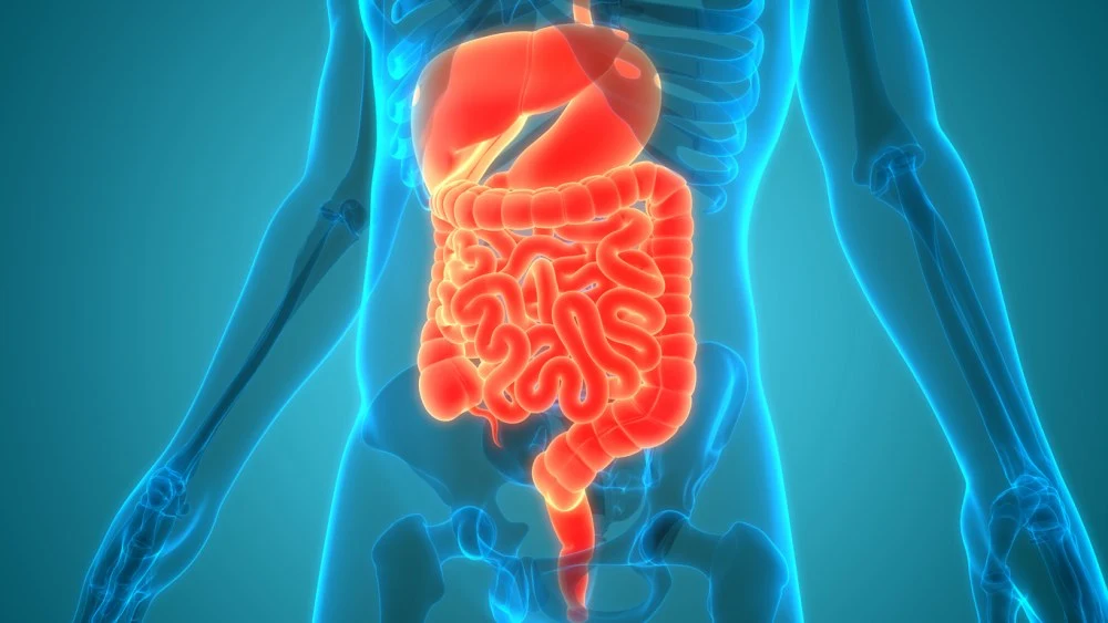
Effective imaging is essential in managing luminal Crohn’s disease (CD), a chronic inflammatory bowel condition marked by unpredictable relapses and remissions. The European Society of Gastrointestinal and Abdominal Radiology (ESGAR) has issued practice recommendations to guide radiologists in diagnosing and monitoring luminal CD, with a focus on cross-sectional imaging modalities such as Magnetic Resonance Enterography (MRE), Intestinal Ultrasound (IUS) and Computed Tomography (CT). These guidelines support clinical decision-making through structured assessment and standardised reporting, with imaging playing a central role in the “treat-to-target” management strategy.
Cross-Sectional Imaging in Diagnosis and Phenotyping
Timely diagnosis and accurate disease phenotyping are crucial for tailoring CD treatment and improving patient outcomes. CD presents with heterogeneous symptoms, and its diagnosis requires integration of clinical, biochemical, histological and imaging data. Cross-sectional imaging techniques—particularly MRE and IUS—have emerged as preferred first-line investigations for suspected small bowel disease. MRE, in particular, offers superior performance in evaluating the extent and distribution of ileal disease, while IUS is sensitive and well tolerated, making it a useful tool for both diagnosis and follow-up.
Must Read: Imaging Techniques for Crohn Disease and Ulcerative Colitis
These modalities provide insights beyond the mucosa, allowing assessment of full bowel wall thickness and extra-enteric complications, which endoscopy cannot visualise. Imaging also helps distinguish between the inflammatory, stricturing and penetrating subtypes of CD. Each subtype carries different therapeutic implications, so phenotyping based on imaging is vital. CT, although effective, is generally reserved for acute or emergency scenarios due to concerns over radiation exposure.
Imaging Indicators and Disease Activity Assessment
A core responsibility of radiologists is to assess disease activity by identifying imaging signs of inflammation. ESGAR recommendations emphasise standardised terminology and structured reporting to support multidisciplinary management. Validated indicators of active disease include bowel wall thickening, mural oedema, perimural inflammation, ulceration and hypervascularity. These parameters are identifiable on both MRE and IUS. For example, mural oedema appears as high signal on fat-suppressed T2-weighted MRE and may disrupt mural stratification on IUS. Additional signs such as altered motility, lymphadenopathy and hypervascularity on Doppler also contribute to the activity profile.
Activity scoring systems, such as the Magnetic Resonance Index of Activity (MaRIA) and various ultrasound-based scores, further standardise assessment. While these tools are more common in research settings, they can improve objectivity in clinical reporting. In practice, a combination of these validated markers allows radiologists to differentiate active from chronic disease and evaluate the severity and distribution of inflammation across the bowel.
Assessing Treatment Response and Identifying Complications
The primary goal of treatment is to control inflammation and prevent irreversible bowel damage. ESGAR supports the use of MRE and IUS for assessing treatment response and monitoring disease progression over time. Radiologists are expected to classify response according to four categories: transmural remission, significant transmural response, stable disease and progressive disease. Transmural remission, the most favourable outcome, involves complete normalisation of mural and perimural parameters. However, this is relatively uncommon and achieved in a minority of patients.
Imaging is also essential for identifying complications such as strictures and penetrating disease. Penetrating complications manifest as fistulae or abscesses, while strictures result from fibrosis and muscle hypertrophy, sometimes accompanied by inflammation. Defining these phenotypes accurately informs the choice of medical, surgical or endoscopic interventions. MRE and IUS are both effective in detecting strictures and assessing their severity. Features such as bowel wall thickening, luminal narrowing and pre-stenotic dilation should be documented. The presence of mixed disease—coexisting inflammation and fibrosis—adds complexity and must be clearly communicated.
Radiologists are integral to the multidisciplinary care of patients with Crohn’s disease, from initial diagnosis to long-term management. ESGAR’s recommendations promote best practices in cross-sectional imaging, advocating the use of MRE and IUS as primary tools for evaluating disease activity, phenotyping and monitoring treatment response. Structured reporting using standard terminology enhances communication and supports informed decision-making. Imaging not only confirms inflammation but also guides intervention by identifying complications and assessing the effectiveness of therapies. These recommendations underscore the importance of imaging in achieving the goals of treat-to-target strategies and improving long-term outcomes in luminal CD.
Source: European Radiology
Image Credit: iStock


