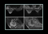
Molecular breast imaging (MBI) is a nuclear medicine technique using technetium 99m (99mTc) sestamibi to image areas of high mitochondrial activity and blood flow in the breast. It is used in various clinical settings, including supplemental screening for dense breast tissue, assessing response to chemotherapy, and as an alternative when MRI is contraindicated. MBI has an incremental cancer detection rate of 7.7–8.8 per 1000 women screened and is less expensive and more tolerable for some patients compared to MRI. Standard evaluation of MBI-detected lesions includes diagnostic mammography or targeted ultrasound (US). However, 15% of lesions are not identifiable with these methods, necessitating further assessment with breast MRI and possibly an MRI-guided biopsy, which can be costly and complex. MBI-guided biopsy presents a cost-effective alternative, improving patient care and health equity. Earlier MBI systems, such as the single-detector breast-specific gamma imaging (BSGI) and the dual-detector MBI systems, had FDA-approved biopsy capabilities but faced limitations due to long procedure times and the current unavailability of biopsy accessories. A novel method for MBI-guided biopsy using a dual-detector system aims to provide a more efficient and feasible approach for performing vacuum-assisted core needle biopsies of MBI-detected lesions.
Prospective Study Design and Procedure Overview
This prospective study, approved by the Mayo Clinic Institutional Review Board, evaluated the feasibility of a novel MBI-guided biopsy system from July 2022 to June 2023. Women aged 25 or older with visible breast findings on any imaging modality were eligible. The study included participants with previously biopsied benign lesions to compare pathology results. Exclusions were for pregnancy, recent lactation, younger age, ipsilateral breast implants, recent breast surgery, or scheduled sentinel lymph node procedures. The study utilised the Stereo Navigator MBI Accessory biopsy unit, an upgrade to the dual-head LumaGem MBI system. The process began with quality assurance checks and a higher dose of 99mTc sestamibi to reduce imaging times. Imaging was done in two orientations, and radiologists selected target lesions for biopsy.
If no biopsy was warranted, follow-up recommendations were based on initial findings. For biopsy, the breast was compressed, and the lesion's position was confirmed using imaging overlays. The upper detector was retracted, and lesion depth was calculated through triangulation. The biopsy procedure involved anaesthetizing the area, making an incision, and using a vacuum-assisted core biopsy needle. Continuous imaging confirmed lesion removal. Specimens were imaged to ensure radiotracer uptake, and additional samples were taken if necessary. A biopsy clip was placed, and a postprocedure mammogram documented the clip placement. Radiology-pathology concordance was assessed post-biopsy.
Participant Outcomes and Procedural Insights
This study, conducted from July 2022 to June 2023, included 21 women (mean age 50.6 years) and aimed to evaluate the feasibility of MBI-guided biopsy. Of these, 17 successfully completed the procedure, while 4 did not undergo biopsy due to non-visualization or reduced lesion size. Lesions biopsied included invasive ductal carcinoma, fibroadenomas, pseudoangiomatous stromal hyperplasia, and fibrocystic changes. No major complications occurred, though one participant developed a hematoma and another had a vasovagal reaction.
Participant characteristics and procedural details, including lesion size, breast thickness, compression force, and procedure time, were recorded. The average lesion size was 17 mm, with an average procedure time of 55 minutes, which reduced to 36 minutes when excluding pre-biopsy imaging. The study documented the number of core biopsy samples and preferred depth calculation method, which was triangulation in all cases.
Reasons for cancelled biopsies included non-visualization or reduced radiotracer uptake. Follow-up imaging showed no subsequent cancer diagnosis in these cases. Overall, the MBI-guided biopsy procedure was deemed feasible and safe, with diagnostic pathology results concordant with imaging findings.
Feasibility, Safety, and Clinical Integration
This study represents the first report of a novel MBI-guided core needle biopsy technique, which uses a retractable upper detector and two methods of lesion depth measurement. The study demonstrated that this MBI-guided biopsy technique is feasible and well tolerated, with no major complications reported. The total procedure time averaged 55 minutes, decreasing to 36 minutes when excluding pre-biopsy imaging.
Key advantages of MBI-guided biopsy include familiar patient positioning, ability to perform specimen imaging during the procedure, and no contraindications related to implants or body habitus, making it accessible to a wider range of patients. The safety profile of 99mTc sestamibi is favourable, with very low rates of adverse reactions. Additionally, MBI-guided biopsy offers a cost-effective alternative to MRI-guided biopsy, with a national average cost of $500 compared to $1000–$3500 for MRI.
Challenges include potential technical difficulties with posterior, subareolar, or superficial lesions, and the absence of a verification rod, which might necessitate further studies to evaluate the need for this step. The study also used a higher radiotracer dose than routine clinical imaging to facilitate shorter imaging times and accurate biopsy targeting.
The study had several limitations, including the small sample size and inclusion of benign lesions, which may not represent the pathology distribution in a population with suspicious lesions. The cancellation rate of 19% for biopsies was higher than previous reports, attributed to the broad inclusion criteria. Future studies are needed to validate these findings and assess the rate of biopsy cancellations further.
The MBI-guided biopsy system using dual-head MBI was feasible, well tolerated, and showed potential for increased integration into clinical practice, enhancing care for patients undergoing MBI.
Source & Image Credit: Radiolog - Imaging Cancer


