Chronic obstructive pulmonary disease (COPD) is a major global cause of death and disability, with substantial impact on health systems and patients’ quality of life. Early diagnosis is critical, as timely intervention can prevent the irreversible damage associated with disease progression. Functional small airway disease (fSAD) represents an intermediate stage between normal lung tissue and emphysema, occurring in the early course of COPD. Detecting fSAD before the onset of extensive emphysematous change can guide prevention strategies and reduce disease burden.
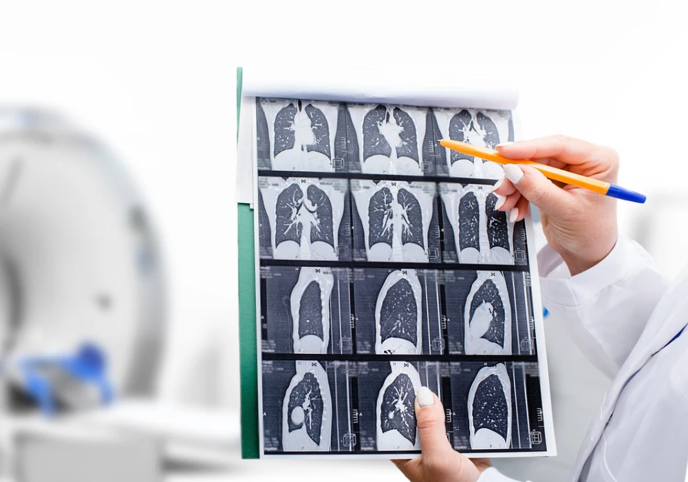
The standard method for evaluating fSAD is parametric response mapping (PRM), a CT-based technique that classifies lung regions into normal, emphysematous or gas-trapping areas by comparing co-registered inspiratory and expiratory scans. However, this requires paired CT imaging, which is rarely recommended in routine clinical practice due to concerns over radiation exposure. The ability to perform accurate PRM evaluation using only a single inspiratory scan could allow for earlier detection of small airway disease, reduce patient radiation dose and support broader use of this diagnostic approach in clinical care.
Generative Modelling for PRM Prediction
To address this need, two deep learning models were developed to predict PRM using only inspiratory chest CT: a predictive model (P-PRM) and a generative model (G-PRM). The P-PRM model applied an end-to-end voxel-wise classification method to assign each lung voxel to a lesion category directly from the inspiratory image. This was implemented using UNet and Attention UNet backbones, optimised with DiceFocal loss to account for class imbalance.
The G-PRM approach followed the principles of traditional PRM calculation. It began by generating a synthetic expiratory scan from the inspiratory CT using a biphase deformable registration process, which aligned structural features before applying intensity transformation to match the expiratory phase. This method allowed fine alignment between inspiratory and generated expiratory images, producing realistic paired scans suitable for PRM analysis. Three image generation techniques were evaluated: a naïve generative method mapping directly from inspiratory to expiratory images, a residual learning scheme predicting the difference between phases, and a generative adversarial network (GAN) approach. The naïve generative method achieved the best results, with the highest structural similarity to real expiratory images and the most accurate PRM outcomes.
The final PRM maps were derived using established attenuation thresholds, classifying voxels into normal lung, fSAD or emphysema. This process mirrors conventional PRM generation, enabling the model’s output to be interpreted in the same way as standard dual-phase imaging while avoiding the second scan.
Performance and Validation
The model development dataset included 308 participants, with a median age of 67 years. These were split into training, validation and first internal test sets. Additional internal test sets were used to assess performance in routine check-up patients, those with comorbid or confounding lung conditions, and a cohort stratified by pulmonary function test (PFT) results.
An external test set from another institution evaluated cross-centre generalisability.
In the first internal test set, the generative model outperformed the predictive model across all metrics. For fSAD detection, G-PRM achieved a sensitivity of 86.3% compared to 38.9% for P-PRM, with AUC improving from 0.70 to 0.86. For emphysema detection, sensitivity rose from 68.7% to 79.6% and AUC from 0.74 to 0.90. These results reflected the generative model’s ability to better capture fine-grained pathological patterns despite class imbalance and diffuse lesion distribution.
Validation on the second internal test set produced AUC values of 0.84 for fSAD, 0.64 for emphysema and 0.97 for normal lung tissue, with an overall accuracy of 80.7%. In the third set, which included patients with conditions such as pneumonia, lung cancer and interstitial lung disease, the model maintained high performance with an accuracy of 89.4% and AUC of 0.83 for fSAD.
The fourth internal test set allowed performance assessment across different PFT-defined groups, including healthy individuals, PRISm patients and COPD GOLD stages I to IV. The model achieved its highest accuracy in the PRISm group at 90.1%, with an AUC of 0.88 for fSAD. This group is of particular interest as it may represent individuals at elevated risk for COPD who do not yet meet spirometric diagnostic thresholds.
External testing on 45 patients from a separate institution confirmed generalisability, with an accuracy of 79.9% and AUC values of 0.85 for fSAD, 0.58 for emphysema and 0.94 for normal lung. The ability to maintain performance across different scanners, acquisition protocols and patient populations supports the model’s potential for wide-scale deployment.
Must Read: Interstitial Lung Abnormalities: Insights from Routine CT Scans
Clinical Implications and Future Directions
The generative deep learning model demonstrates that accurate PRM can be achieved using only a single inspiratory CT scan, offering a clinically practical and radiation-sparing alternative to conventional dual-phase imaging. By preserving the interpretability of PRM maps and generating realistic synthetic expiratory scans, the approach allows radiologists and clinicians to visualise and quantify disease with outputs aligned to established workflows.
In practice, this capability could enable earlier identification of small airway disease, guiding preventive interventions such as smoking cessation and targeted treatment before significant lung damage occurs. The strong performance in the PRISm subgroup suggests potential for risk stratification in populations not yet diagnosed with COPD, opening opportunities for early disease management.
From a technical perspective, the success of G-PRM reflects its ability to address two key challenges: the low prevalence of lesion voxels relative to normal tissue, and the diffuse distribution of early pathological changes. By simulating the transformation between inspiratory and expiratory phases, the model effectively preserves subtle lesion patterns that voxel-wise classification models may miss. The use of dynamic thresholding during PRM generation also provides flexibility, allowing clinicians to adjust sensitivity and specificity according to clinical priorities.
Future research will aim to extend validation through larger, prospective multicentre studies to confirm generalisability and reproducibility. Model refinements may include improved registration algorithms to minimise loss of fine structural detail and integration of real PRM maps as additional supervision to further enhance accuracy. Addressing the small but noted risk of overpredicting fSAD in some normal or mild COPD cases will also be a priority.
A generative deep learning approach using a single inspiratory chest CT scan can accurately detect functional small airway disease, achieving performance comparable to conventional dual-phase PRM while substantially reducing radiation exposure. It has shown robustness across diverse patient groups, disease severities and imaging conditions, with particular value for identifying at-risk individuals before COPD diagnosis. Its interpretability, adaptability and potential for integration into CT workflows make it a promising tool for improving early detection and management of small airway disease in clinical practice.
Source: Radiology: Artificial intelligence
Image Credit: iStock


