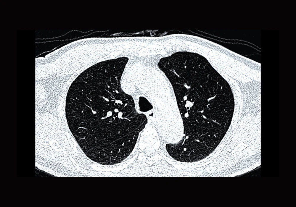
Low-dose CT screening aims to find lung cancer earlier, but the value of artificial intelligence as an independent reader depends on how reliably it flags scans with risk-dominant nodules and assigns the correct nodule type. An evaluation of baseline screening in a Chinese general population compared reconstruction choices to see how they affect AI behaviour. The work assessed kernels and slice thickness or interval against a radiologist consensus reference and focused on the risk-dominant nodule that guides management. Kernel selection altered scan-level sensitivity and nodule type agreement, whereas a modest change in slice thickness and interval made little difference. Aligning AI inputs with the kernel used by radiologists improved consistency, which matters for routine screening workflows.
Population and Imaging Approach
Three hundred consecutive participants from the NELCIN-B3 programme in Tianjin were included after excluding cases with missing or poor-quality reconstructions. The mean age was 61.2 years, 53.0% were women, and 35.6% were current or former smokers with a mean 22.7 pack-years. All scans used a 128-detector row CT in a low-dose protocol. Reconstructions comprised a medium-soft kernel at 1 mm (D45f/1 mm), a sharp kernel at 1 mm (B80f/1 mm), and a soft kernel at 2 mm (B30f/2 mm). To isolate thickness effects under the same kernel, a 2-mm series was also created from D45f data by software reformation.
Internal link: Limits of AI in Pulmonary Nodule Classification
AI analysis used a commercial deep learning system that detects, segments and classifies nodules. The pipeline had been trained on a public dataset, generated candidates with convolutional networks, and reduced false positives with a second stage. Segmentation combined intensity thresholds with a deformable model. Classification produced four types: noncalcified solid, part-solid, nonsolid or calcified. Outputs included nodule location and size, including solid components.
Two radiologists read all scans independently and reached a consensus reference. They searched for nodules using maximum intensity projections under D45f with 10-mm slabs, then used multiplanar reformations under B80f at 1-mm slices for volumetry. The risk-dominant nodule was defined as the noncalcified solid or part-solid nodule with the largest solid component whenever any such nodule measured at least 30 mm³. Scans without a qualifying solid component were recorded as having no risk-dominant nodule. Performance was measured at scan level for the presence of a risk-dominant nodule and at case level for detection of the reference risk-dominant nodule and agreement in type classification.
Kernel Effects on Detection
Radiologist agreement on the presence of a risk-dominant nodule was almost perfect. AI agreement with the reference depended strongly on kernel selection. With the medium-soft kernel at 1 mm, agreement was moderate to substantial and scan-level sensitivity was clearly higher than with the sharp kernel at the same thickness. Specificity differences between these two 1-mm settings were small. When thickness and interval changed from 1.0/0.7 mm to 2.0/1.0 mm under the same medium-soft kernel, AI sensitivity and specificity were not meaningfully altered. Performance under the two 2-mm reconstructions showed no important advantage for either kernel in scan-level detection, reinforcing that the large shift came from kernel choice at thin slices rather than from the thickness change within the tested range.
On a case basis, AI detected the reference risk-dominant nodule in a high proportion of cases across all reconstructions. The rate did not differ significantly between kernels or between the two D45f thickness settings. When the reference risk-dominant nodule was detected, it matched the largest AI nodule only in a little over half to about two thirds of cases depending on reconstruction. This alignment defined the subset used for comparing type classification.
Examples highlighted systematic blind spots that were independent of kernel. Very large lesions beyond a preset volume threshold were treated as masses by the algorithm and were not flagged as nodules. Nodules attached to vessels or the pleura could also go undetected across reconstructions despite meeting the risk-dominant definition by the reference. These edge cases show that kernel selection improves general performance but does not remove all detection limits.
Kernel Effects on Nodule Type
For matched nodules, kernel selection had a marked effect on type classification. Agreement with the reference was high with the medium-soft kernel at 1 mm and fell sharply with the sharp kernel at 1 mm. At 2 mm, agreement remained higher with the medium-soft option than with the soft kernel. There was no meaningful difference between 1-mm and 2-mm series under the medium-soft kernel for type classification, which again points to kernel choice as the main driver within the tested thickness range.
Patterns of error differed by kernel. With the sharp kernel, noncalcified solid nodules were frequently mislabelled as calcified, shifting cases away from the risk-dominant category. With the soft 2-mm kernel, noncalcified solid nodules were more often misclassified as nonsolid. Both patterns matter because the presence and type of the risk-dominant nodule determine scan-level categorisation and follow-up. Using the same kernel as the radiologists improved alignment in type, likely because image texture and edges were represented in a similar way, which made the AI outputs more consistent with human assessment.
These effects have practical consequences in screening pathways. Scans without a risk-dominant nodule usually fall into routine interval follow-up. A sharp kernel that reduces sensitivity and distorts type classification increases the chance of labelling a scan as negative when a risk-dominant nodule is present. In contrast, using the radiologist-aligned medium-soft kernel supported higher agreement without sacrificing specificity.
Limitations and Generalisability
Several factors temper interpretation. The reference was based on radiologist consensus rather than histopathology. Slice thicknesses above 2 mm were not examined. An upper volume threshold in the AI could exclude large but clinically relevant lesions from nodule consideration. The evaluation used one commercial system in a single centre with an automatic workflow, so external validation with other algorithms and settings is needed. Even so, the size and direction of the kernel effect on both scan-level sensitivity and type classification were consistent across the tested parameters.
Kernel selection is the dominant technical choice shaping AI behaviour in low-dose CT lung screening. A medium-soft kernel aligned with radiologist reading improved scan-level sensitivity and substantially increased agreement in nodule type, whereas a sharp kernel reduced both. Changing from thin to 2-mm slices under the same medium-soft kernel did not meaningfully alter performance, suggesting that kernel alignment should be the priority for programmes seeking dependable AI support. Emphasising the right kernel can help integrate AI as an independent reader with greater confidence in day-to-day screening.
Source: European Radiology
Image Credit: iStock


