Researchers in South Korea previously reported that diffusion-weighted magnetic resonance imaging (DW-MRI) could be used to predict neurologic outcomes before targeted temperature management (TTM) after return of spontaneous circulation (ROSC) from cardiac arrest. In a new study, the same team of researchers compared the efficacy of brain computed tomography (CT) and DW-MRI to predict neurologic outcome before TTM in comatose cardiac arrest survivors.
You might also like: Drones May Improve Response Times for Out-Of-Hospital Cardiac Arrests
Their new finding suggests that DW-MRI is potentially useful for early prediction of neurologic outcome (i.e., before TTM) in cardiac arrest patients. The researchers explain that the combination of grey matter to white matter ratio (GWR) on brain CT and that on DW-MRI, rather than on each modality alone, appears to improve the sensitivity for predicting neurologic outcome after ROSC from cardiac arrest. The new study is published in the journal Resuscitation.
The Korean team performed a retrospective study of cardiac arrest patients treated with TTM. The brain CT and DW-MRI were both obtained before TTM. The researchers analysed GWR on the brain CT and the presence of high signal intensity on DW-MRI, alone or in combination, to predict poor neurologic outcome.
Of 47 comatose cardiac arrest patients treated with TTM, 39 patients with brain CT and DW-MRI data were included in the analysis. Median time from the ROSC to the brain CT and DW-MRI was 90 minutes (52–150) and 175 minutes (118–240), respectively. There was no significant difference in predicting poor neurologic outcome between average GWR (area under the curve [AUC] 0.891, sensitivity/specificity 78.8%/100%) and DW-MRI (AUC 0.894, sensitivity/specificity 75.8%/100%). The combination of average GWR and DW-MRI (AUC 0.939, sensitivity/specificity 87.9%/100%) improved the prediction of poor neurologic outcome rather than each one alone and in other combinations.
"To the best of our knowledge, this is the highest AUC and sensitivity of the GWR test in predicting neurologic outcome among the published studies. The most important reasons for variations in the results of GWR on brain CT in predicting neurologic outcome are the ability of the interpreter and the measurement errors," the researchers say.
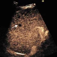
Cortical laminar necrosis of the brain may be identified within hours of the anoxic-ischaemic event, and DW-MRI offers a good visualisation of laminar necrosis and other characteristic signs of hypoxic injury and unsurpassed promptness, according to the research team. Cerebral cortical diffusion abnormalities are known to be associated with poor neurologic outcome after cardiopulmonary arrest. On DWI sequences, MRI changes after global anoxic-ischaemic brain injury due to cardiac arrest appear as a hyper-intensity in cortical areas or basal ganglia after ROSC, the team says.
The study has some important limitations. Specifically, researchers note that the generalisability of their findings is limited because this study was a retrospective study performed at a single centre with small number of patients. Therefore, large-scale multicentre studies should be conducted for generalisation of the results.
Source: Resuscitation
Image Credit: U.S. Navy photo by Photographer's Mate 2nd Class Timothy Comerford
References:
Jeon, Chi Heon et al. (2017) Comparison of brain computed tomography and diffusion-weighted magnetic resonance imaging to predict early neurologic outcome before target temperature management comatose cardiac arrest survivors. Resuscitation. doi.org/10.1016/j.resuscitation.2017.06.021
Latest Articles
cardiac arrest, computed tomography, Neurologic Outcome, diffusion-weighted magnetic resonance imaging
In a new study, researchers compared the efficacy of brain computed tomography (CT) and DW-MRI to predict neurologic outcome before TTM in comatose cardiac arrest survivors.