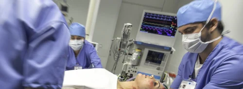
This article provides an overview of transoesophageal echocardiography training, programme development, feasibility and impact on the diagnosis and treatment of critically ill patients.
Recently clinicians at our centre managed one of the most critical patient emergencies. An elderly woman (Patient A) presented to our emergency department (ED) by ambulance with cardiac arrest of unknown aetiology. Information on her medications was initially unavailable and history provided by her family was nonspecific: she had experienced generalised malaise, diarrhoea and poor oral intake for several days. Patient A also had a history of hypertension, obesity, hypothyroidism, dyslipidaemia and atrial fibrillation.
Upon arrival, pulseless electrical activity (PEA) was detected and she was endotracheally intubated while cardiopulmonary resuscitation (CPR) and advanced cardiac life support (ACLS) protocols were initiated. After one round of CPR and resuscitative medications, Patient A remained in PEA. ACLS guidelines advocate for continued resuscitation while simultaneously considering and treating potentially reversible causes of the cardiac arrest (Sayre et al. 2010). Transthoracic echocardiography (TTE) was attempted, but it provided no meaningful information due to ongoing CPR.
This case illustrates several challenges clinicians face during the resuscitation of patients in cardiac arrest and how our team uses transoesophageal echocardiography (TEE) to reliably obtain high-quality images, guide decision-making and intra-arrest procedures, and monitor response - without interrupting lifesaving chest compressions. This article provides an overview of TEE training, programme development, feasibility and impact on the diagnosis and treatment of critically ill patients at London Health Sciences Centre, a tertiary care center consisting of two hospitals in Ontario, Canada with 50 intensive care unit (ICU) beds at two sites and two emergency departments (EDs) with 140,000 combined annual visits. Lifesaving applications of TEE in the ED and ICU are also reviewed.
A 97% Success Rate in Answering High-Stakes Clinical Questions in Critically Ill Patients
A TEE probe was inserted without difficulty to reveal a midoesophageal four-chamber view with no evidence of pericardial effusion or signs of cor pulmonale suggestive of pulmonary embolism (PE). Ultrasound-guided central venous catherisation (CVC) revealed a high-risk relationship between the internal jugular vein and carotid artery. The high risk of arterial cannulation was minimised by using TEE to confirm proper venous guidewire placement with a midoesophageal bicaval view.
ACLS and European Resuscitation Council guidelines have recently endorsed echocardiography in cardiac arrest resuscitation, as have earlier guidelines from cardiology and anaesthesiology societies (Cheitlin et al. 2003; Thys et al. 2010; Link et al. 2015; Soar et al. 2015). In 2017, the American College of Emergency Physicians (ACEP) published the first guidelines endorsing the use of TEE by emergency physicians (EPs), reporting that in up to 50% of cases, TTE provides inadequate images in critically ill patients and is even more challenging to perform in those receiving CPR (Fair et al. 2018). Moreover, TTE also risks interrupting chest compressions for more than the ten seconds advised in the ACLS guidelines, potentially leading to worse neurological outcomes in cardiac arrest patients.
The ACEP guidelines report that TEE “provides the logical solution to these limitations, given its ability for continuous image acquisition both during compressions and during pulse checks, its reliably excellent image quality and its lack of interference with chest compressions or other procedures needed during cardiac arrest.” Indeed, TEE’s superior image quality in nearly all circumstances and expanded diagnostic scope due to its indwelling location millimetres behind the heart have shown a very high success rate in answering high-stakes clinical questions in severely ill patients [97% for TEE versus 38% for TTE] (Vignon et al. 1994). For cardiac arrest resuscitation, the ACEP guidelines cite the following benefits of TEE:
- TEE provides a valuable adjunct for diagnosing myocardial infarction, PE, pericardial effusion and hypovolaemia as causes of the arrest.
- Anaesthesia literature has demonstrated that TEE can reliably identify the cause of the arrest in up to 86% of cases, offering potential advantages in being able to confidently guide such treatment decisions as the use of thrombolysis, vasopressors, intravenous fluid or blood administration, or pericardiocentesis.
- TEE offers immediate, real-time feedback on the response to any intervention, such as visualisation of coordinated contractility after defibrillation or improvement in contractility after administering epinephrine.
- TEE provides immediate assessment of the quality of chest compressions. The 2015 ACLS guidelines advise a specific compression depth of 5 to 6 centimetres during CPR - a goal that can be hard to clinically evaluate without TEE.
- TEE can also assist with the proper placement of intra-aortic balloon pumps, transvenous pacemakers and other resuscitative devices.
A Safe, Clinically Influential and Easy-to-Learn Technique
With Patient A remaining in a persistent PEA rhythm of five beats per minute, transcutaneous pacing was attempted, but failed due to her body habitus. Placement of a 5F balloon-directed transvenous pacemaker was performed under direct visualisation using TEE, which proved very valuable in the context of difficult electrical capture. Once capture was achieved, good blood pressure was confirmed. The return of circulation post-capture enabled us to rule out acute coronary syndrome and PE as causes of the arrest.
Studies by our team and other investigators reveal that TEE is safe, feasible and clinically influential in a range of emergency and critical care scenarios. In an ICU case series published by our team, 80% of intensivist-performed TEE studies at our centre have resulted in proposed changes in management (Arntfield et al. 2018), versus 60% of TTE studies as published in an earlier study at our centre (Alherbish et al. 2015). The TEE study analysed findings from 274 consecutive TEE examinations performed by 38 operators, with the most common indications being haemodynamic instability (45.2%), assessment for infective endocarditis (22.2%), poor TTE windows (20.1%), and cardiac arrest (20.1%). Some studies carried more than one indication. All TEE examinations were safely performed and produced interpretable images, with a 100% success rate for probe insertion (84% on the first pass) and no mechanical complications.
Our study found that TEE is a powerful diagnostic tool that can answer both advanced and basic questions essential for the daily care of the critically ill, including the determination of shock aetiology, preload sensitivity, procedural support (extracorporeal membrane oxygenation cannulation, central venous catheter insertion, cardioversion) and monitoring of haemodynamic interventions. In our study, we found that about two-third of the TEE exams addressed basic questions, using a limited number of views. In the 42% of cases in which TTE was performed prior to TEE, unsatisfactory image quality led to TEE in half of these cases.
Given the compelling evidence of TEE’s superior performance in critically ill patients, and the availability of TEE-compatible portable ultrasound machines and high-fidelity simulators for training, broad dissemination of TEE training to EPs is now a realistic consideration. Our critical care team developed and evaluated a novel focused TEE examination tailored for use in the ED by EPs (Arntfield et al. 2015). TEE-naïve EPs were invited to participate in a didactic and simulation-based workshop where they learned how to obtain views from four vantage points (mid-oesophageal four-chamber, mid-oesophageal long axis, transgastric short-axis and bicaval views). After the training, their skills were assessed on a high-fidelity simulator and a six-week follow-up session assessed skill retention, demonstrating that EPs can successfully perform the focused TEE examination and retained those skills six weeks later.
Other investigators have reported that although use of TEE takes practice, since the user must learn to manipulate the probe remotely, mastering this skill is actually easier with TEE than with TTE, because the probe is well positioned simply by being in the oesophagus (Mayo et al. 2015). Unlike TTE, TEE is generally uninfluenced by positive pressure ventilation, obesity, emphysema, surgical dressings or wounds, and obstacles on the chest, such as defibrillator pads, or ongoing CPR. Many of the image planes and views generated by TTE and TEE are similar, differing only in how they are projected onto the screen. Moreover, the techniques used for evaluation of the cardiac anatomy and function are identical.
Impact of Focused TEE Examinations in the Emergency Department
Remarkably, after the return of paced circulation, Patient A began to move purposefully and required sedation. Time from the initial cardiac arrest until successful pacemaker capture was about 45 minutes. Ultimately, the cause of her cardiac arrest was found to be hyperkalaemia. Information on her medications was obtained, leading to a diagnosis of acute kidney injury from a diarrhoeal illness in the context of use of one of her medications.
In a recent retrospective study of all ED TEE examinations performed by EPs at our centre between February 1, 2013 and January 31, 2015, this safe, minimally invasive tool imparted a diagnostic influence in 78% of cases and impacted therapeutic decisions in 67%. In all cases, probe insertion was successful and the views obtained were determinate in 98% of cases. Focused TEE exams demonstrated the most promise in patients who were intubated and had undifferentiated shock or cardiac arrest (Arntfield et al. 2016).
Patient A’s case, which has been more fully described elsewhere (Arntfield et al. 2014), powerfully demonstrates the value of point-of-care TEE in rapid evaluation for reversible causes of arrest, guiding invasive procedures during emergency scenarios and providing continuous, real-time anatomic monitoring without pauses in lifesaving chest compressions to acquire images, as is necessary with TTE. Use of TEE during her resuscitation was like watching a live TV show in which we could actually see the heart of a patient who had been brought in with absent vital signs start to beat again. After correction of her potassium level, Patient A was no longer pacemaker dependent and was discharged to her home with full neurological and functional recovery six days later.
When we telephoned Patient A to follow up on the case, we expected her to sound weak and fatigued after her near-fatal illness. Instead, she sounded joyful and full of life. “I was playing with my grandkids,” she announced. In the background, we could hear the excited voices of children clamouring for Grandma to return to their game. It is countless stories like this that continue to inspire us to use point-of-care TEE in our ICUs and EDs to uphold and improve the standard of care for critically ill patients, provide diagnostic certainty in emergency scenarios, including cardiac arrest, and guide lifesaving procedures, even if the use of TTE is impossible. The goal of our TEE programme is simple: to use the best available technology and techniques to help our sickest patients get back in the game.
Key Points
- TEE provides a valuable adjunct for diagnosing myocardial infarction, pulmonary embolism, pericardial effusion and hypovolaemia as causes of the arrest.
- TEE can reliably identify the cause of the arrest in up to 86% of cases.
- TEE offers immediate, real-time feedback on the response to any intervention.
- TEE provides immediate assessment of the quality of chest compressions.
- TEE can also assist with the proper placement of intra-aortic balloon pumps, transvenous pacemakers and other resuscitative devices.
Alherbish A, Priestap F, Arntfield R (2015) The introduction of basic critical care echocardiography reduces the use of diagnostic echocardiography in the intensive care unit. J Crit Care, 30(6):1419.e7-1419.e11.
Arntfield R, Lau V, Landry Y et al. (2018) Impact of Critical Care Transesophageal Echocardiography in Medical–Surgical ICU Patients: Characteristics and Results From 274 Consecutive Examinations. J Intensive Car Med, Sep 6: 885066618797271.
Arntfield R, Pace J, McLeod S, Granton J, Hegazy A, Lingard L (2015) Focused transesophageal echocardiography for emergency physicians—description and results from simulation training of a structured four-view examination. Crit Ultrasound J, 7:10.
Arntfield R, Pace J, Hewak M et al. (2016) Focused transesophageal echocardiography by emergency physicians is feasible and clinically influential: observational results from a novel ultrasound program. J Emerg Med, 50:286-294.
Arntfield RT, Millington SJ, Wu E (2014) An Elderly Woman That Presents With Absent Vital Signs. Chest, 146:3146-e159.
Cheitlin MD, Armstrong WF, Aurigemma GP et al. (2003) ACC/AHA/ASE 2003 Guideline Update for the Clinical Application of Echocardiography: Summary Article: A Report of the American College of Cardiology/American Heart Association Task Force on Practice Guidelines (ACC/AHA/ASE Committee to Update the 1997 Guidelines for the Clinical Application of Echocardiography). Circulation, 108:1146-1162.
Fair J, Mallin M, Mallemat H, Zimmerman J, Arntfield R et al. (2018) Transesophageal Echocardiography: Guidelines for Point-of-Care Applications in Cardiac Arrest Resuscitation. Annals of Emergency Medicine, 71(2):201-207.
Link MS, Berkow LC, Kudenchuk PJ, et al. (2015) Part 7: adult advanced cardiovascular life support: 2015 American Heart Association guidelines update for cardiopulmonary resuscitation and emergency cardiovascular care. Circulation, 132(18 suppl 2):S444-S464.
Mayo PH, Narasimhan M, Koenig S. (2015) Critical Care Transophageal Echocardiography. Chest, 148(5):1323-1332.
Sayre MR, Koster RW, Botha M et al. (2010) Adult Basic Life Support Chapter Collaborators. Part 5: adult basic life support: 2010 international consensus on cardiopulmonary resuscitation and emergency cardiovascular care science with treatment recommendations . Circulation, 122(16)(suppl 2 ):S298 -S32.
Soar J, Nolan JP, Böttiger BW et al. (2015) European Resuscitation Council Guidelines for Resuscitation 2015: Section 3. Adult advanced life support. Resuscitation, 95:100-147.
Thys DM, Brooker RF, Cahalan MK, et al. (2010) Practice guidelines for perioperative transesophageal echocardiography. An updated report by the American Society of Anesthesiologists and the Society of Cardiovascular Anesthesiologists Task Force on Transesophageal Echocardiography. Anesthesiology, 112:1084-1096.
Vignon P, Mentec H, Terré S, et al. (1994) Diagnostic accuracy and therapeutic impact of transthoracic and transesophageal echocardiography in mechanically ventilated patients in the ICU. Chest, 106(6):1829–1834.


