Without Primary Acute Brain Injury
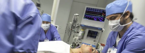
This review focuses on the current experience with clinically available neuromonitoring techniques in critically ill patients at risk for neurological compromise, but without overt acute brain injury (ABI).
The field of neuromonitoring has grown rapidly over the past 30 years, which has helped improve pathophysiological understanding, clinical care and outcomes for patients with primary ABI, including traumatic brain injury (TBI), subarachnoid haemorrhage (SAH), hypoxic-ischaemic injury and ischaemic or haemorrhagic stroke. The main goals of neuromonitoring are to better understand a patient’s cerebral physiology, provide early detection of neurological worsening or cerebral dysfunction to avoid progression to irreversible neurological injury, and assist with neuroprognostication. This is accomplished through a combination of serial neurological examinations, neuroimaging studies, and continuously monitoring different neurophysiological parameters. Numerous expert opinion and evidence-based reviews on the role of multimodality neuromonitoring in ABI have been published, including a
consensus statement on multimodality monitoring in neurocritical care from the Neurocritical Care Society and the European Society of Intensive Care Medicine (Stocchetti et al. 2013; Le Roux et al. 2014).
Most research and clinical practice in neuromonitoring has focused on patients with primary ABI, particularly TBI and SAH, and occurred in specialised neurocritical or neurotrauma care units (NCCUs). There is also a large population of critically ill patients without primary neurologic disease or ABI, who are at high risk for cerebral injury from their underlying disease process, systemic complications, or medical therapies, e.g. cardiac arrest, severe sepsis from extracranial sources, endocrinopathies such as diabetic ketoacidosis and hyper-/ hypo-thyroidism, renal and hepatic failure, rheumatologic conditions like haemophagocytic lymphohistiocytosis, and haematological abnormalities, including leukaemias and other neoplasms. The pathophysiology of secondary ABI in these patients is complex given the heterogeneous aetiologies and the numerous physiological cascades that can contribute to cerebral injury. Like ABI patients, these high-risk patients may benefit from neurophysiological and neurodiagnostic techniques to detect the antecedents of neurologic insults, thereby identifying a therapeutic window when neuroprotective or neurorestorative interventions will have the greatest likelihood of preventing and minimising irreversible brain injury.
Clinical Components of Multimodality Neuromonitoring
The traditional cornerstone of clinical neuromonitoring is the performance of serial bedside neurological examinations. The main components of the examination in a critical care environment comprise an assessment of the patient’s mental status, including consciousness and awareness, cranial nerves and gross motor abilities. The
Glasgow Coma Scale (GCS) is the most widely used scale for characterising a patient’s degree of consciousness (Teasdale and Jennett 1974). Although it was originally developed to assess consciousness in TBI patients, its use has since expanded, becoming the most commonly used tool to communicate global neurological status for all ICU patients. It has been tested widely and has reasonable intra-rater and inter-rater reliability, although accuracy is greater amongst more experienced providers (Rowley and Fielding 1991). Concerns exist about its accuracy and usefulness in intubated patients and those receiving sedatives, analgesics and neuromuscular blocking agents (Kornbluth and Bhardwaj 2011). A newer scale to measure the degree of consciousness is the
Full Outline of UnResponsiveness (FOUR) score, which includes assessments of eye and motor responsiveness similar to the GCS, and adds tests for brainstem reflexes (pupil and corneal reflexes) and respiratory pattern (Wijdicks et al. 2005; Iyer et al. 2009). Both the GCS and FOUR score measures perform well at predicting in-hospital mortality and functional outcome (Wijdicks et al. 2011), although the utility of detecting subtle neurological changes in critically ill patients with these scales is unclear. Delirium is also a common manifestation of neurologic dysfunction in critically ill patients, and routine use of screening tools like the
Confusion Assessment Method for the ICU (CAM-ICU) or the
Intensive Care Delirium Screening Checklist (ICDSC) is recommended (Barr et al. 2013). Abnormalities detected while performing serial neurological examinations, whether on coma or delirium assessment scales, brainstem reflexes or detailed clinical examination could indicate the presence or evolution of ABI, and further investigation with neuroimaging or other diagnostic modalities may then be required.
Neuroimaging
Computerised tomography (CT) and magnetic resonance imaging (MRI) are the most frequently used modalities to diagnose or exclude intracranial pathology in critically ill patients. They are typically performed in response to new or changing neurologic signs or symptoms, and are not routinely used as screening tools. CT is commonly used to evaluate critically ill patients with an acute change in neurological status, and can readily detect pathology like intracranial haemorrhage, hydrocephalus, and cerebral oedema, which may require emergency medical or surgical intervention. Portable CT imaging is now readily available and provides high quality images; because no patient transport is required this reduces the risk of transport-associated brain insults and complications (Peace et al. 2010; Swanson et al. 2010). MRI has greater anatomical resolution and a variety of specialised imaging sequences that provide detailed maps demonstrating subtle areas of infarction or haemorrhage, integrity of white matter tracts and quantitative information about cerebral physiology, perfusion, and metabolism. These images, however, are acquired at single time points during evolving systemic and cerebral disease processes. While these images can provide insights into disease pathophysiology, explain clinical symptoms and provide prognostic information, they always should be related to — and combined with — continuous neuromonitoring techniques and the clinical evaluation.
Electrophysiology
Continuous electroencephalography (cEEG) monitoring provides essential information while monitoring critically ill patients at risk for ABI. Continuous EEG is the only means to detect electrical (i.e. nonconvulsive) seizures and monitor response to anticonvulsant therapy. Seizures can be a manifestation of an occult cerebral insult, and in some circumstances they can aggravate existing brain injury. Current guidelines recommend cEEG monitoring in ICU patients without primary ABI who have “unexplained impairment of mental status or unexplained neurological deficits” to evaluate for nonconvulsive seizures (Claassen et al. 2013). Seizures occur in about 10% of ICU patients without primary ABI, with severe sepsis, renal failure and hepatic failure being the most significant risk factors (Oddo et al. 2009). Nonconvulsive seizures exclusively occurred in 67% of cases (Oddo et al. 2009). ICU patients with unexplained neurological dysfunction are typically monitored with cEEG for 24-48 hours (Gavvala et al. 2014). Seizures in critically ill patients are associated with poor outcomes and increased mortality. Additionally, EEG abnormalities (e.g. abnormal background frequencies, triphasic waves, etc.) have been associated with mortality in septic patients (Young et al. 1992). Although only currently used in patients with ABI from subarachnoid haemorrhage, characteristic trends on cEEG correlate with delayed cerebral ischaemia from vasospasm have been used at the bedside to supplement clinical data (Rathakrishnan et al. 2011; Vespa et al. 1997). Modules that quantitatively process EEG waveforms and graphically display information (e.g. compressed spectral array and colour density spectral array EEG) can also be used at the bedside for seizure detection and to monitor for patterns of cerebral dysfunction from causes like ischaemia.
Unlike EEG, evoked potentials are typically recorded as isolated snapshots and generally used to help estimate prognosis in select cases, e.g. bilateral absent N20 peaks after cardiac arrest rather than guide therapy (Wijdicks et al. 2006; Carter and Butt 2001). In addition, it is difficult to maintain the stability of EPs to perform continuous monitoring (Fossi et al. 2006).
Intracranial Pressure, Cerebral Blood Flow and Cerebral Autoregulation
Although intracranial pressure (ICP) and its consequences are discussed frequently in critical care patients, it is rarely measured directly (e.g. ventriculostomy or intraparenchymal monitor) in patients without ABI. Noninvasive techniques to estimate ICP include measuring optic nerve sheath diameter with ultrasound (Ohle et al. 2015; Rajajee et al. 2011), two-depth transcranial doppler (TCD) insonation of the ophthalmic artery (Ragauskas et al. 2012), TCD-derived pulsatility index (Bouzat et al. 2010), and computationally from the relationship between the arterial blood pressure and the middle cerebral artery (MCA) flow velocity waveforms (Schmidt et al. 2003). While these methodologies have shown encouraging results, they are mostly intermittent measures aimed at screening patients with clinical suspicion for increased ICP. The TCD-based approaches have the potential for continuous monitoring, but require constant adjustment of probe placement and have technical issues relating to device compatibility. In one study using a noninvasive TCD-based technique, ICP measurements in septic patients did not increase above 20 mmHg after fluid resuscitation. Calculated CPP levels below 60, however, were associated with increased neuronal injury, as assessed by S-100ß levels (Pfister et al. 2008a).
In circumstances when the clinical history, neurological examination and neuroimaging suggest cerebral oedema and that ICP increases may lead to ABI, invasive ICP monitors can be placed to accurately assess ICP and guide medical therapy. This has been done for patients with acute liver failure (Mohsenin 2013; Blei et al. 1993), diabetic ketoacidosis (Srinivasan et al. 2012) and drug intoxications (Marklund et al. 2007). In these patients, continuous ICP monitoring is more clinically useful than intermittent measurements.
Cerebrovascular pressure reactivity is the mechanism through which cerebral vessels protect the brain against inappropriate cerebral blood flow (CBF) changes, irrespective of acute changes in blood pressure or cerebral perfusion pressure (CPP). This is accomplished by the brain’s intrinsic ability to dynamically adjust cerebrovascular tone or resistance. Dysregulation of this system can contribute to cerebral insults by several mechanisms, including ischaemia, hyperaemia, hypoxia, increased ICP and cerebral energy dysfunction. Cerebrovascular autoreactivity can be assessed in a dynamic fashion by interrogating the relationship between arterial blood pressure (ABP) or CPP and measures of cerebral blood flow or volume. Techniques to measure CBF used in this manner are MCA flow velocity via TCD, ICP, brain tissue oxygen (PbtO2) and near-infrared spectroscopy (NIRS) (Zweifel et al. 2014).
Impaired cerebrovascular autoreactivity has been demonstrated after ABI and as a component of other systemic disease processes like severe sepsis (Taccone et al. 2010; Pfister et al. 2008b), liver failure (Macias-Rodriguez et al. 2015), and diabetic ketoacidosis (Ma et al. 2014). Some of the neurological complications in patients with these conditions may result in part from loss of cerebral autoregulation. There are currently no medical therapies to improve impaired cerebral autoregulation, although evidence exists that autoregulation may be optimised for an individual patient within a specified range of ABPs or CPPs and carbon dioxide levels (Taccone et al. 2010; Aries et al. 2012; Steiner et al. 2002). Additionally, sedative medications may influence cerebrovascular reactivity and this effect may be modulated by patient age and underlying disease process (Kadoi et al. 2008; Hinohara et al. 2005).
Cerebral Metabolism and Brain Oxygenation
Numerous pathological cascades that involve impaired brain oxygenation and cerebral metabolism exist. These can precipitate or exacerbate neurological insult in critically ill patients. Several monitoring modalities are available to interrogate brain oxygen, including direct measurement of brain tissue oxygen tension in a specific brain region (PbtO2), global cerebral oxygen delivery via jugular bulb venous oxygen saturation (SjvO2) and noninvasive cerebral oximetry in frontal brain regions with NIRS (Barazangi and Hemphill 2008; Maloney-Wilensky and Le Roux 2010; Stocchetti et al. 2013). There is limited experience with these techniques in patients without primary ABI, although the potential information learned about cerebral oximetry and metabolic function could prove helpful to predict, manage and prevent neurologic compromise in patients at high risk for brain injury.
A few studies have used these techniques to evaluate patients with sepsis with neurological symptoms (Oddo and Taccone 2015; Taccone et al. 2013). In septic patients with altered mental status, SjvO2 and flow velocity in the MCA with TCD were measured during a dobutamine challenge. While CBF and oxygen delivery increased with dobutamine, oxygen consumption did not change (Berre et al. 1997). Another study found poor agreement between CBF estimates derived from TCD and NIRS (Toksvang et al. 2014). NIRS has potential wide applicability to examine for cerebral compromise, e.g. noninvasive evaluation of cerebral autoregulation in septic patients (Steiner et al. 2009), to assess cognition-related abnormalities in brain function in patients with mild hepatic encephalopathy (Nakanishi et al. 2014), to evaluate cerebral hyperaemia during treatment for diabetic ketoacidosis (Glaser et al. 2013), and to monitor cerebral function during volume resuscitation in dehydrated patients (Hanson et al. 2009). The best results however may be in neonates, and further study is required in adults.
Cerebral metabolism can be assessed through the sampling of the brain’s extracellular fluid with surgically implanted cerebral microdialysis catheters. This technique provides an evaluation of regional cerebral bioenergetics; abnormal concentrations of certain cerebral metabolites (i.e. lactate, pyruvate, glucose, glutamate) can indicate evolving energy failure, hypoxia or ischaemia, or an imbalance between aerobic and anaerobic metabolism (Hutchinson et al. 2014; 2015). Most clinical experience with microdialysis is with ABI, and TBI and SAH in particular. However, it has been used in monitoring platforms of fulminant hepatic failure (Hutchinson et al. 2006; Tofteng et al. 2002). The ability of cerebral microdialysis to interrogate the brain’s neurochemistry may increase our understanding of the pathophysiology of other systemic diseases that have neurological complications, like septic encephalopathy, diabetic ketoacidosis, toxic exposures, and assist in the critical care management of these patients by providing targets to protect against cerebral energy failure. For example, in TBI patients, tight glycaemic control (80-120 mg/dL) with aggressive insulin therapy was associated with reduced cerebral glucose concentrations and worse cerebral energy crisis (Oddo et al. 2008; Vespa et al. 2012). Both high and low cerebral glucose have been associated with poor outcome in TBI. It is unclear what the optimal range for brain glucose is, what the relationship between serum and brain glucose concentrations in a metabolically stressed brain is, and how generalisable these findings are to other types of neurological insults (Hutchinson et al. 2015). However, knowledge about brain glucose may help guide therapy. In addition, understanding metabolism has shown that alternative fuels such as lactate may be beneficial in select patients (Oddo et al. 2012; Bouzat et al. 2014).
Conclusions and Future Directions
A major challenge to prevent brain injury in critically ill patients is that the progression from neuronal dysfunction to permanent injury typically proceeds undetected through a critical period when neuroprotective or neurorestorative interventions are likely to be effective. The goal of multimodality monitoring is to integrate signals in real time from a variety of technologies to provide bedside clinicians with a metric of the relative health or dysfunction of the brain before, during and after this critical period. Physicians can then use this information to guide individualised and goal-directed therapy to help prevent and mitigate further neurological injury. The integrative capabilities of bioinformatics platforms allow for the rapid synthesis and display of this data in a format that provides clinicians reliable information that can be used to target therapies (Hemphill et al. 2011).
Selecting the appropriate components of a multimodality platform must take into account the underlying pathophysiology of the relevant disease processes and potential mechanisms of cerebral injury. For example, neurological sequelae in patients with severe sepsis occur in multiple patterns including ischaemic strokes, vasogenic oedema and white matter abnormalities (Stubbs et al. 2013). Areas of perfusional ischaemia may result from impaired cerebral autoregulation or systemic hypotension, while other areas of injury may be the result of deleterious inflammatory mediated processes like endothelial cell swelling, microvascular dysfunction, alterations in blood-brain barrier permeability, and neurotransmitter imbalance. Thus, measuring cerebral autoregulation or microdialysis may detect actionable precursors to cerebral dysfunction in this population, whereas measuring ICP may be less helpful, because rarely is there enough cerebral oedema to increase ICP.
Choosing the most appropriate systemic and neurophysiological monitor is particularly important for patients who are at high risk for brain insult, but have not yet sustained brain injury. Currently, strategies to manage these patients are reactive in nature, with physicians responding to changes in neurological exam or physiological variables. Multimodality neuromonitoring has the capability to provide high-resolution real-time physiological data that, when computationally integrated and synthesised, can facilitate proactive measures to detect and correct neurophysiological derangements to avoid neurological compromise (Wartenberg et al. 2007). Furthermore, this paradigm will allow for an enriched understanding of the brain’s response to systemic pathological states, and help design targeted treatment strategies that are mechanistically derived rather than empiric in nature. Overall, this rapidly evolving technology will provide physicians with additional information to promote brain health and avoid neuronal insult in critically ill patients.
See Also:
Sedation in Acute Brain Injury: Less is More?
Aries MJ, Czosnyka M, Budohoski KP et al. (2012)
Continuous determination of optimal cerebral perfusion pressure in traumatic
brain injury. Crit Care Med, 40(8): 2456-63.
Barazangi N,
Hemphill JC (2008) Advanced cerebral monitoring in neurocritical care. Neurol
India, 56: 405-14.
Barr J, Fraser GL,
Puntillo K et al. (2013) Clinical practice guidelines for the management of
pain, agitation, and delirium in adult patients in the intensive care unit.
Crit Care Med, 41(1): 263-306.
Berré J, De
Backer D, Moraine JJ et al. (1997) Dobutamine increases cerebral blood flow
velocity and jugular bulb hemoglobin saturation in septic patients. Crit Care
Med, 25(3): 392-8.
Blei AT,
Olafsson S, Webster S et al. (1993) Complications of intracranial pressure
monitoring in fulminant hepatic failure. Lancet, 341(8838): 157-8.
Bouzat P,
Francony G, Fauvage B et al. (2010) Transcranial Doppler pulsatility index for
initial management of brain-injured patients. Neurosurg, 67: E1863-4; author
reply E1864.
Bouzat P,
Magistretti PJ, Oddo M (2014) Hypertonic lactate and the injured brain: facts
and the potential for positive clinical implications. Intesive Care Med, 40(6):
920-1.
Carter BG, Butt
W (2001) Review of the use of somatosensory evoked potentials in the prediction
of outcome after severe brain injury. Crit Care Med, 29: 178-86.
Claassen J,
Taccone FS, Horn P et al. (2013) Recommendations on the use of EEG monitoring
in critically ill patients: consensus statement from the neurointensive care
section of the ESICM. Intensive Care Med, 39(8): 1337-51.
Fossi
S, Amantini A, Grippo A et al. (2006) Continuous
EEG-SEP monitoring of severely brain injured patients in NICU: methods and
feasibility. Neurophysiol Clin, 36(4):
195-205.
Gavvala J, Abend
N, Laroche S et al. (2014) Continuous EEG monitoring: a survey of
neurophysiologists and neurointensivists. Epilepsia, 55(11): 1864-71.
Glaser NS,
Tancredi DJ, Marcin JP et al. (2013) Cerebral hyperemia measured with near
infrared spectroscopy during treatment of diabetic ketoacidosis in children. J
Pediatr, 163(4): 1111-6.
Hanson SJ,
Berens RJ, Havens PL et al. (2009) Effect of volume resuscitation on regional
perfusion in dehydrated pediatric patients as measured by two-site
near-infrared spectroscopy. Pediatr Emerg Care, 25(3): 150-3.
Hemphill JC,
Andrews P, De Georgia M (2011) Multimodal monitoring and neurocritical care
bioinformatics. Nat Rev Neurol, 7(8): 451-60.
Hinohara H,
Kadoi Y, Takahashi K et al. (2005) Differential effects of propofol on
cerebrovascular carbon dioxide reactivity in elderly versus young subjects. J
Clin Anesth, 17(2): 85-90.
Hutchinson P, O'Phelan
K, Participants in the International Multidisciplinary Consensus Conference on
Multimodality Monitoring (2014) International multidisciplinary consensus
conference on multimodality monitoring: cerebral metabolism. Neurocrit Care, 21
Suppl 2: S148-58.
Hutchinson PJ,
Gimson A, Al-Rawi PG et al. (2006) Microdialysis in the management of hepatic encephalopathy.
Neurocrit Care, 5(3): 202-5.
Hutchinson PJ,
Jalloh I, Helmy A, Carpenter KL, Rostami E, Bellander BM, Boutelle MG, Chen JW,
Claassen J, Dahyot-Fizelier C, Enblad P, Gallagher CN, Helbok R, Hillered L, Le
Roux PD, Magnoni S, Mangat HS, Menon DK, Nordström CH, O'Phelan KH, Oddo M,
Perez Barcena J, Robertson C, Ronne-Engström E, Sahuquillo J, Smith M,
Stocchetti N, Belli A, Carpenter TA, Coles JP, Czosnyka M, Dizdar N, Goodman
JC, Gupta AK, Nielsen TH, Marklund N, Montcriol A, O'Connell MT, Poca MA,
Sarrafzadeh A, Shannon RJ, Skjøth-Rasmussen J, Smielewski P, Stover JF,
Timofeev I, Vespa P, Zavala E,
Ungerstedt U (2015) Consensus statement from the 2014 International
Microdialysis Forum. Intensive Care Med, 41(9): 1517-28.
Iyer VN,
Mandrekar JN, Danielson RD et al. (2009) Validity of the FOUR score coma scale
in the medical intensive care unit. Mayo Clin Proc, 84(8): 694-701.
Kadoi Y, Saito S,
Kawauchi C et al. (2008) Comparative effects of propofol vs dexmedetomidine on
cerebrovascular carbon dioxide reactivity in patients with septic shock. Br J
Anaesth, 100(2): 224-9.
Kornbluth J,
Bhardwaj A (2011) Evaluation of coma: a critical appraisal of popular scoring
systems. Neurocrit Care, 14(1): 134-43.
Le Roux P, Menon
DK, Citerio G et al. (2014) Consensus summary statement of the International
Multidisciplinary Consensus Conference on Multimodality Monitoring in
Neurocritical Care : a statement for healthcare professionals from the
Neurocritical Care Society and the European Society of Intensive Care Medicine.
Intensive Care Med, 40(9): 1189-209.
Ma L, Roberts
JS, Pihoker C et al. (2014) Transcranial Doppler-based assessment of cerebral
autoregulation in critically ill children during diabetic ketoacidosis
treatment. Pediatr Crit Care Med, 15(8): 742-9.
Macías-Rodríguez
RU, Duarte-Rojo A, Cantú-Brito C et al. (2015) Cerebral haemodynamics in
cirrhotic patients with hepatic encephalopathy. Liver Int, 35(2): 344-52.
Maloney-Wilensky
E, Le Roux P (2010) The physiology behind direct brain oxygen monitors and
practical aspects of their use. Childs Nerv Syst, 26(4): 419-30.
Marklund N,
Enblad P, Ronne-Engström E (2007) Neurointensive care management of raised
intracranial pressure caused by severe valproic acid intoxication. Neurocrit
Care, 7(2): 160-4.
Mohsenin V (2013)
Assessment and management of cerebral edema and intracranial hypertension in
acute liver failure. J Crit Care, 28(5): 783-91.
Nakanishi H,
Kurosaki M, Nakanishi K et al. (2014) Impaired brain activity in cirrhotic
patients with minimal hepatic encephalopathy: Evaluation by near-infrared
spectroscopy. Hepatol Res, 44(3): 319-26.
Oddo M, Carrera
E, Claassen J et al. (2009) Continuous electroencephalography in the medical
intensive care unit. Crit Care Med, 37(6): 2051-6.
Oddo M, Schmidt
JM, Carrera E, Badjatia N, Connolly ES, Presciutti M, Ostapkovich ND, Levine JM,
Le Roux P, Mayer SA (2008) Impact of tight glycemic control on cerebral glucose
metabolism after severe brain injury: a microdialysis study. Crit Care Med, 36(12):
3233-8.
Oddo M, Levine J, Frangos S, Maloney-Wilensky
E, Carrera E, Daniel R , Magistretti PJ, Le Roux P (2012)
Brain lactate metabolism in humans with subarachnoid haemorrhage. Stroke,
43(5):1418-21.
Oddo M, Taccone
FS (2015) How to monitor the brain in septic patients? Minerva Anestesiol, 81(7):
776-88.
Ohle R, Mcisaac
SM, Woo MY et al. (2015) Sonography of the Optic Nerve Sheath Diameter for
Detection of Raised Intracranial Pressure Compared to Computed Tomography: A
Systematic Review and Meta-analysis. J Ultrasound Med, 34(7): 1285-94.
Peace
K, Maloney E, Frangos S, MacMurtrie E,
Shields E, Hujcs M, Levine J, Kofke A, Yang W, Le Roux P (2010a) The
use of a portable head CT scanner in the ICU. J Neurosci Nursing,
42(2): 109-16.
Pfister D,
Schmidt B, Smielewski P et al. (2008a) Intracranial pressure in patients with
sepsis. Acta Neurochir Suppl, 102: 71-5.
Pfister D,
Siegemund M, Dell-Kuster S et al. (2008b) Cerebral perfusion in
sepsis-associated delirium. Crit Care, 12(3): R63.
Ragauskas A,
Matijosaitis V, Zakelis R et al. (2012) Clinical assessment of noninvasive
intracranial pressure absolute value measurement method. Neurology, 78(21):
1684-91.
Rajajee V,
Vanaman M, Fletcher JJ et al. (2011)
Optic nerve ultrasound for the detection of raised intracranial pressure.
Neurocrit Care, 15(3): 506-15.
Rathakrishnan R,
Gotman J, Dubeau F et al. (2011) Using continuous electroencephalography in the
management of delayed cerebral ischemia following subarachnoid hemorrhage.
Neurocrit Care, 14(2): 152-61.
Rowley G,
Fielding K (1991) Reliability and accuracy of the Glasgow Coma Scale with
experienced and inexperienced users. Lancet, 337(8740): 535-8.
Schmidt B, Czosnyka
M, Raabe A et al. (2003) Adaptive noninvasive assessment of intracranial
pressure and cerebral autoregulation. Stroke, 34(1): 84-9.
Srinivasan S,
Benneyworth B, Garton HJ et al. (2012) Intracranial pressure/cerebral perfusion
pressure-targeted management of life-threatening intracranial hypertension
complicating diabetic ketoacidosis-associated cerebral edema: a case report.
Pediatr Emerg Care, 28(7): 696-8.
Steiner LA,
Czosnyka M, Piechnik SK et al. (2002) Continuous monitoring of cerebrovascular
pressure reactivity allows determination of optimal cerebral perfusion pressure
in patients with traumatic brain injury. Crit Care Med, 30(4): 733-8.
Steiner LA,
Pfister D, Strebel SP et al. (2009) Near-infrared spectroscopy can monitor
dynamic cerebral autoregulation in adults. Neurocrit Care, 10(1): 122-8.
Stocchetti N, Le
Roux P, Vespa P et al. (2013) Clinical review: neuromonitoring - an update.
Crit Care, 17(1): 201.
Stubbs DJ,
Yamamoto AK, Menon DK (2013) Imaging in sepsis-associated
encephalopathy--insights and opportunities. Nat Rev Neurol, 9(10): 551-61.
Swanson
E, Mascitelli J, Stiefel M, MacMurtrie E,
Levine J, Kofke WA, Yang W, Le Roux P (2010b) The effect of
patient transport on brain oxygen in comatose patients. Neurosurgery,
66(5): 925-32.
Taccone FS,
Castanares-Zapatero D, Peres-Bota D et al. (2010) Cerebral autoregulation is
influenced by carbon dioxide levels in patients with septic shock. Neurocrit
Care, 12(1): 35-42.
Taccone FS,
Scolletta S, Franchi F, Donadello K et al. (2013) Brain perfusion in sepsis.
Curr Vasc Pharmacol, 11(2): 170-86.
Teasdale G,
Jennett B (1974) Assessment of coma and impaired consciousness. A practical
scale. Lancet, 2(7872): 81-4.
Tofteng F,
Jorgensen L, Hansen BA et al. (2002) Cerebral microdialysis in patients with
fulminant hepatic failure. Hepatology, 36(6): 1333-40.
Toksvang LN,
Plovsing RR, Petersen MW et al. (2014) Poor agreement between transcranial
Doppler and near-infrared spectroscopy-based estimates of cerebral blood flow
changes in sepsis. Clin Physiol Funct Imaging, 34(5): 405-9.
Vespa P,
Mcarthur DL, Stein N et al. (2012) Tight glycemic control increases metabolic
distress in traumatic brain injury: a randomized controlled within-subjects
trial. Crit Care Med, 40(6): 1923-9.
Vespa PM, Nuwer
MR, Juhasz C et al. (1997) Early detection of vasospasm after acute
subarachnoid hemorrhage using continuous EEG ICU monitoring. Electroencephalogr
Clin Neurophysiol, 103(6): 607-15.
Wartenberg KE,
Schmidt JM, Mayer SA (2007) Multimodality monitoring in neurocritical care.
Crit Care Clin, 23(3): 507-38.
Wijdicks EF,
Bamlet WR, Maramattom BV et al. (2005) Validation of a new coma scale: The FOUR
score. Ann Neurol, 58(4): 585-93.
Wijdicks
EF, Hijdra A, Young GB et al. (2006) Practice
parameter: prediction of outcome in comatose survivors after cardiopulmonary
resuscitation (an evidence-based review): report of the Quality Standards
Subcommittee of the American Academy of Neurology. Neurology,
67(2): 203-10.
Wijdicks EF,
Rabinstein AA, Bamlet WR et al. (2011) FOUR score and Glasgow Coma Scale in
predicting outcome of comatose patients: a pooled analysis. Neurology, 77(1):
84-5.
Young GB, Bolton
CF et al. (1992) The electroencephalogram in sepsis-associated encephalopathy.
J Clin Neurophysiol, 9(1): 145-52.
Zweifel C, Dias
C, Smielewski P et al. (2014) Continuous
time-domain monitoring of cerebral autoregulation in neurocritical care. Med
Eng Phys, 36(5): 638-45.


