A new mathematical model has been developed by a team of scientists at John Hopkins led by cardiologist and biomedical engineer Hiroshi Ashikaga, M.D, PhD. The model can measure and digitally map the beat-sustaining electrical flow between heart cells. The approach has been described in the Journal of the Royal Society Interface.
The team believes that this new model could form a blueprint for more precise imaging tests that would be able to capture cell-to-cell communication. They will also be able to pinpoint the tiny clusters of cells at the epicentre of complex, life-threatening arrhythmias. Such a precise imaging approach would enable targeted and minimally invasive treatments that could eliminate rhythm-disrupting hotspots in the heart's electrical system.
The approach used by the scientists is inspired by information theory and is built on the premise that cell-to-cell interaction follows a classic model of communication that consists of source, transmitter and receiver. It translates cellular conversations into digital form that can be read and imaged by a computer, enabling clinicians to spot breakdowns in communication that form the epicentres of dangerous rhythm disturbances.
"Successful arrhythmia treatment depends on correctly identifying the epicentre of the malfunction," Ashikaga says. "We cannot begin to develop such precision-targeted therapies without understanding the exact nature of the malfunction and its precise location. This new model is a first step toward doing so."
The basic concept behind this model is that heart muscle cells act as analog-to-digital converters and take up information from their surroundings, convert and interpret this information and transmit the message to neighbouring cells. This capturing and quantifying of information can help catch aberrant signs as they trigger electrical firestorms that cause the heart to beat abnormally and compromise its ability to pump blood.
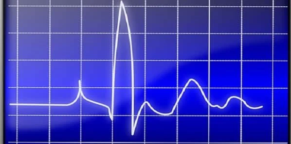
Electrocardiograms provide limited information and are most helpful in diagnosing the type of arrhythmia rather than the exact cellular origin of the rhythm disturbance. However, this new approach creates computer representations of normal and abnormal heartbeats, ranging from simpler benign arrhythmias to dangerous rhythms. When using this model, the scientists digitised the electric flow converting the electrical signals transmitted by cells into bits. They then measured how much information was generated, transmitted and received during normal and abnormal heart rhythms and plotted this information on a 2D map to create an image of the arrhythmia.
This mathematical model could help pinpoint the origin of dangerous arrhythmias and could lead to new therapies as well as improve the precision of surgical ablation.
Source: John Hopkins Medicine
Image Credit: Pixabay

