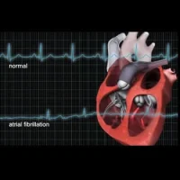

Researchers from Johns Hopkins have found that specific altered function in the left atrium may signal stroke risk in those with atrial fibrillation, and, possibly, those without it. The findings have been reported in the Journal of the American Heart Association.
The researchers performed sophisticated motion studies by using an imaging technique that combines standard MRI scans with a motion-tracking software that analyses cardiac movement. The research team analysed records of 169 patients aged 49 to 69 with atrial fibrillation who had cardiac MRIs before undergoing a minimally invasive procedure to ablate small sections of the heart tissue. Eighteen of these patients had suffered minor or major strokes prior to the ablations. Using the sophisticated imaging technique, they compared images of the hearts of the patients who had suffered strokes with those who had not.
They observed that the left atria of patients who had suffered strokes had reduced ability to empty out blood into the lower portion of the heart as compared to patients without strokes. They also found that the left atria of patients with stroke were bigger and more dilated. Overall, the left atria of patients who'd suffered stroke had worse ability to stretch out and recoil with each heartbeat. These findings show that patients who had suffered strokes had suppressed function and slower blood turnover.
The results of their study suggest that certain features of the heart's upper left chambers seen on heart MRI can could be used to tell apart high risk patients from low-risk patients. The results also cast doubt on the current dogma that the chaotic beating of the upper chambers of the heart during atrial fibrillation fuels blood clot formation that causes stroke. The researchers point out that this view does not explain why many people with atrial fibrillation never have strokes and why many with atrial fibrillation present no evidence of abnormal rhythms within a month before the stroke.
The researchers believe that the suppression function and altered anatomy of the left atrium may play a role in the sluggish blood flow leading to clot formation and precipitating stroke.
"Our observations suggest that altered function in the left atrium of the heart may lead to stroke independently of the heart rhythm disturbance itself," says co-investigator Joao Lima, M.D., professor of medicine and radiology at the Johns Hopkins University School of Medicine and director of cardiovascular imaging at The Johns Hopkins Hospital. "What this means is that people with compromised function in the left upper portion of the heart may be at risk for stroke, with or without atrial fibrillation."
Lead investigator and heart rhythm specialist Hiroshi Ashikaga, M.D., Ph.D., assistant professor of medicine and biomedical engineering at the Johns Hopkins University School of Medicine suggests that maybe atrial fibrillation is not the real culprit and it may be the dysfunction of the left atrium that may be responsible. He indicates that this will be further evaluated in an upcoming study.
Source: Johns Hopkins Medicine
Image Credit: YouTube