Thoracic Insufficiency Syndrome (TIS) is a severe condition where the thorax cannot support normal breathing or lung development, primarily affecting paediatric patients. This syndrome often arises from congenital deformities or malformations in the thoracic spine or ribs, which can significantly impair respiratory function. Proper assessment and monitoring of lung parenchymal aeration are critical for understanding the physiological status of the lungs in patients with TIS, both before and after treatment interventions. Current imaging techniques, such as computed tomography (CT), hyperpolarised gas magnetic resonance imaging (MRI), and single-plane two-dimensional (2D) dynamic MRI (dMRI), face various limitations, including radiation exposure, complexity, and the need for patient cooperation. This recent article published in Radiology: Cardiothoracic Imaging discusses the development and implementation of a novel dMRI technique designed to measure lung aeration, offering a promising alternative to existing methods.
Challenges with Existing Imaging Techniques
Conventional imaging methods, including CT and MRI, have been essential in diagnosing and assessing TIS. However, these techniques present significant challenges that limit their practical application, particularly in paediatric populations. CT scans, while providing detailed images, involve ionising radiation, posing a health risk, especially with repeated exposure. Hyperpolarised gas MRI, though capable of delivering high-resolution images, requires specialised equipment and gases that are not readily available in all clinical settings. Additionally, this technique often necessitates breath-holding or the use of a spirometer to monitor breathing, which can be challenging for young children or patients with severe respiratory compromise.

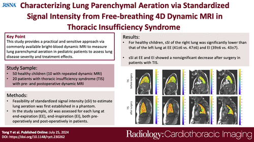
Image Source: Radiology: Cardiothoracic Imaging
Single-plane 2D dMRI, another technique used to assess lung function, provides dynamic imaging of lung movement but lacks the three-dimensional (3D) resolution necessary for comprehensive analysis. Moreover, these methods typically require patient cooperation, such as breath-holding, which is difficult to achieve consistently in paediatric patients. Consequently, there is a need for a more practical, non-invasive, and accurate method for assessing lung aeration in patients with TIS and other respiratory conditions.
The Role of dMRI in Assessing Lung Aeration
To address these challenges, researchers have developed a quantitative dMRI technique that enables the measurement of lung dynamics, including lung volumes and tidal volumes, without the need for breath-holding or specialised gases. This method, known as bright-blood dMRI, utilises a 3-Tesla MRI scanner and true fast imaging with a steady-state precession sequence. It achieves high spatial resolution (approximately 1 × 1 × 6 mm³) with a relatively short acquisition time (11-13 minutes), making it feasible for routine clinical use, particularly in paediatric settings.
The study involved a cohort of 40 healthy children and 20 patients with TIS, who underwent dMRI scans both before and after vertical expandable prosthetic titanium rib (VEPTR) surgical treatment. VEPTR is a U.S. Food and Drug Administration–approved intervention for TIS, involving the implantation of expandable titanium rods that support the spine and ribs, allowing for better lung expansion and growth. The dMRI technique developed in this study measures standardised signal intensity (sSI) values, which are derived from the intensity of MR images and standardised across all patients to reflect specific tissue characteristics. This standardisation allows for accurate comparison of lung aeration across different individuals and time points.
Study Findings and Implications
The study's results revealed significant findings regarding lung aeration in both healthy children and patients with TIS. In healthy children, the sSI values decreased with age, indicating changes in lung aeration associated with normal lung development. For patients with TIS, the study demonstrated that sSI values decreased significantly after VEPTR surgery, suggesting improved lung aeration postoperatively. This decrease in sSI was more pronounced during end inspiration (EI) compared to end expiration (EE), reflecting enhanced lung function during maximal lung expansion.
Additionally, the study included a phantom experiment to validate the accuracy of the dMRI technique. The phantom consisted of seven polyvinyl acetate sponge blocks with varying air content, and the sSI values obtained from the dMRI scans correlated strongly with the actual air content, demonstrating the technique's reliability in measuring lung aeration. The repeatability of the sSI measurements was also assessed by scanning a subset of healthy children twice, with results showing high consistency between scans, further confirming the method's reliability.
The study's findings underscore the potential of this dMRI technique as a valuable tool for assessing lung function in clinical settings. Its ability to provide detailed, 3D images of lung aeration during free breathing, without the need for breath-holding or specialised equipment, makes it particularly advantageous for paediatric patients. The technique's high spatial resolution and short acquisition time also make it practical for routine use, providing clinicians with a powerful tool for monitoring lung function and guiding treatment decisions in patients with TIS and other respiratory conditions.
The development of a quantitative dMRI technique for assessing lung parenchymal aeration represents a significant advancement in managing Thoracic Insufficiency Syndrome and potentially other respiratory disorders. This method offers a non-invasive, practical, and reliable way to measure lung function, overcoming the limitations of traditional imaging techniques. The ability to obtain high-resolution images during free breathing, coupled with the method's high repeatability and accuracy, positions this technique as a valuable tool for both clinical and research applications. Future studies could explore the use of this technique in different patient populations, further refine the imaging protocols, and expand the analysis to more detailed regional assessments within the lungs. This innovative approach holds great promise for improving the diagnosis, treatment planning, and monitoring of lung function in paediatric patients and beyond.
Source: Radiology: Cardiothoracic Imaging
Image Credit: iStock


