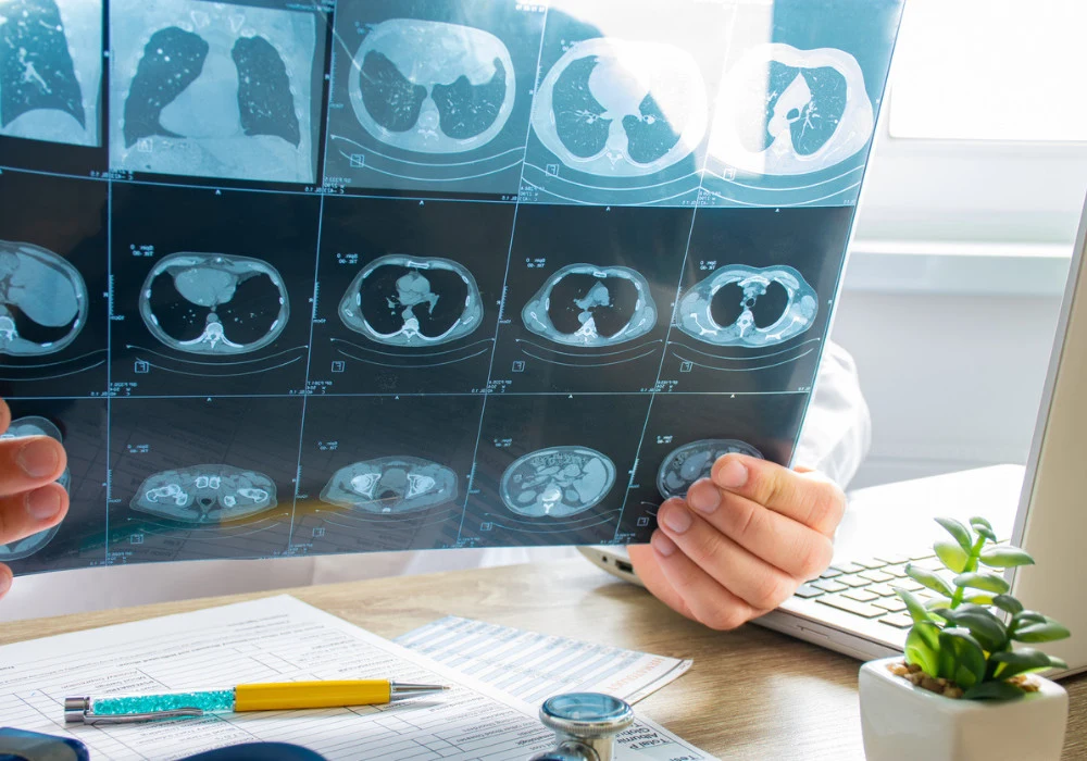
The advent of artificial intelligence (AI) in medical imaging has revolutionised the way radiologists and clinicians assess and interpret diagnostic scans. Chest computed tomography (CT) scans, frequently performed for lung cancer screening, have the potential to reveal critical extrapulmonary findings that go beyond the lungs, including cardiovascular conditions and other abnormalities that can significantly affect patient mortality. Integrating AI tools to detect such findings improves early diagnosis and offers predictive insights into all-cause mortality (ACM). A recent article published in Radiology evaluates AI-based multi-structure segmentation to predict ACM and identify clinically relevant incidental findings in chest CT scans.
AI and Extrapulmonary Findings
Incidental findings on chest CT scans often include significant abnormalities beyond the lungs, such as cardiovascular diseases. In the National Lung Screening Trial (NLST) cohort, extrapulmonary findings were reported in 33.8% of participants, underscoring their clinical importance. The study proposes an AI system capable of analysing 32 structures visible in chest CT scans. Focused on pulmonary findings, this AI can also identify structures like coronary arteries and epicardial adipose tissue (EAT), which indicate cardiovascular risk.
AI-based segmentation tools, such as TotalSegmentator, were used to identify and quantify radiomic features from the segmented structures. This allows for a comprehensive evaluation of cardiovascular conditions, especially coronary artery calcium (CAC), which has been identified as a significant predictor of mortality. Moreover, by integrating radiomic data with demographic factors, the study’s AI models provided clinicians with a robust tool to detect high-risk patients, aiding early intervention and treatment planning.
Predicting All-Cause Mortality Using AI
One of the central goals of the AI tool was to predict ACM over short-term (2-year) and long-term (10-year) periods. During the study, approximately 14% of the 24,401 participants died within a 10-year follow-up period, underscoring the predictive capability of the AI model. The AI system achieved an area under the receiver operating characteristic curve (AUC) of 0.72 for 10-year ACM prediction and 0.71 for 2-year ACM, demonstrating its reliability in assessing mortality risk.
The AI’s ability to predict ACM was largely driven by the importance of coronary artery calcification. In fact, the study showed that participants with higher CAC scores were significantly more likely to experience cardiovascular events leading to death. By focusing on clinically relevant structures and combining demographic data such as age, sex, and smoking history, the AI tool provided a personalised risk score for each patient. This feature is particularly valuable in clinical settings, where timely and accurate risk assessments can inform treatment pathways and potentially save lives.
Impact of AI on Clinical Practice
The integration of AI in medical imaging promises a new era of precision diagnostics. The use of AI-driven multi-structure segmentation enhances the detection of both primary and incidental findings, improving diagnostic accuracy. By identifying extrapulmonary abnormalities such as EAT and CAC, clinicians can better manage patient risk and prioritise follow-up treatments.
The AI system’s ability to predict mortality through multi-structure analysis marks a significant advancement in patient care. Rather than relying solely on radiologists to identify potential risks, AI supplements their expertise with detailed, objective data. For example, while radiologists may focus on primary lung cancer screening, the AI system ensures that cardiovascular risks and other extrapulmonary conditions are not overlooked. This holistic approach leads to more comprehensive patient evaluations, which is particularly crucial for individuals undergoing screening for lung cancer or other high-risk conditions.
The study highlights AI's transformative role in medical imaging, particularly in identifying incidental findings and predicting all-cause mortality. By leveraging multi-structure segmentation and radiomic analysis, AI tools provide clinicians with powerful insights into patient health, enabling earlier interventions and more precise treatment strategies. AI’s integration into routine diagnostic practices will likely lead to significant improvements in patient outcomes, especially in detecting life-threatening conditions that may not be immediately apparent. The future of medical imaging lies in AI’s ability to synthesise vast amounts of data, providing both predictive and explanatory power that enhances clinical decision-making.
Source: Radiology
Image Credit: iStock
References:
Marcinkiewicz AM, Buchwald M, Shanbhag A et al. (2024) AI for Multistructure Incidental Findings and Mortality. Prediction at Chest CT in Lung Cancer Screening. Radiology 312(3): e240541


