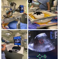
Traditionally, the risk of preterm birth has been evaluated based solely on a woman's history of previous premature deliveries, leaving first-time pregnancies without a reliable risk assessment method. However, a groundbreaking development in the use of Quantitative Ultrasound (QUS) scanning of the cervix during routine pregnancy ultrasounds offers a promising solution. This novel technique significantly enhances the accuracy of predicting preterm birth risks.
Innovative QUS Method Offers Tissue-Level Insights
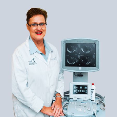
Developed through over twenty years of collaborative research between nursing and engineering experts at the University of Illinois Chicago and the University of Illinois Urbana-Champaign, this innovative method involves the detailed analysis of microstructural changes in the cervix. The research, published in the American Journal of Obstetrics & Gynaecology Maternal-Fetal Medicine, indicates the effectiveness of this ultrasound method from as early as the 23rd week of pregnancy. Professor Emeritus Barbara McFarlin from UIC underscores the technique's ability to provide crucial insights based on tissue characteristics, moving beyond traditional symptomatic indicators like ruptured membranes.
Early Detection of Preterm Birth Risk
In a study with 429 women at the University of Illinois Hospital who went into labour spontaneously, this new method accurately identified first-time pregnancy preterm birth risks. Additionally, in subsequent pregnancies, combining the woman’s delivery history with quantitative ultrasound data offered a more comprehensive risk assessment than using historical data alone. Unlike standard ultrasound imaging that provides visual images, QUS analyses radio frequency data to determine tissue properties. Given that 10-15% of pregnancies are at risk of preterm birth, understanding these underlying factors is vital. Early identification of preterm risk could lead to increased monitoring and timely interventions to delay labour, improving patient outcomes.
Cross-Disciplinary Research for Enhanced Patient Care
The genesis of this research dates back to 2001, initiated by McFarlin during her doctoral studies. Teaming up with Professor Bill O’Brien, an expert in electrical and computer engineering at UIUC, they explored QUS's potential in detecting cervical changes indicative of preterm delivery. The study, backed by the National Institutes of Health’s National Institute of Child Health and Human Development, includes a diverse team of experts. Their goal is to further investigate preventive measures for preterm birth and refine the accuracy of QUS.
The advent of this new ultrasound method heralds a significant leap forward in prenatal care. By enabling early detection of preterm birth risk, particularly in first-time pregnancies, it paves the way for more effective interventions and improved outcomes for both mothers and infants. This research underscores the importance of cross-disciplinary collaboration in advancing medical science and patient care.
Source : AJOG MFM
Image Credit: Barbara McFarlin, lead researcher


