In a ESR/EFSUMB joint session chaired by Caroline Ewertsen (Denmark) and Paul S. Sidhu (UK), lecturers presented the latest advances in small parts multiparametric ultrasound (MPUS) examinations. Maija Radzina (Latvia) concluded the discussions with an outline of future perspectives for AI applications in MPUS.
What is the available evidence of AI in ultrasound?
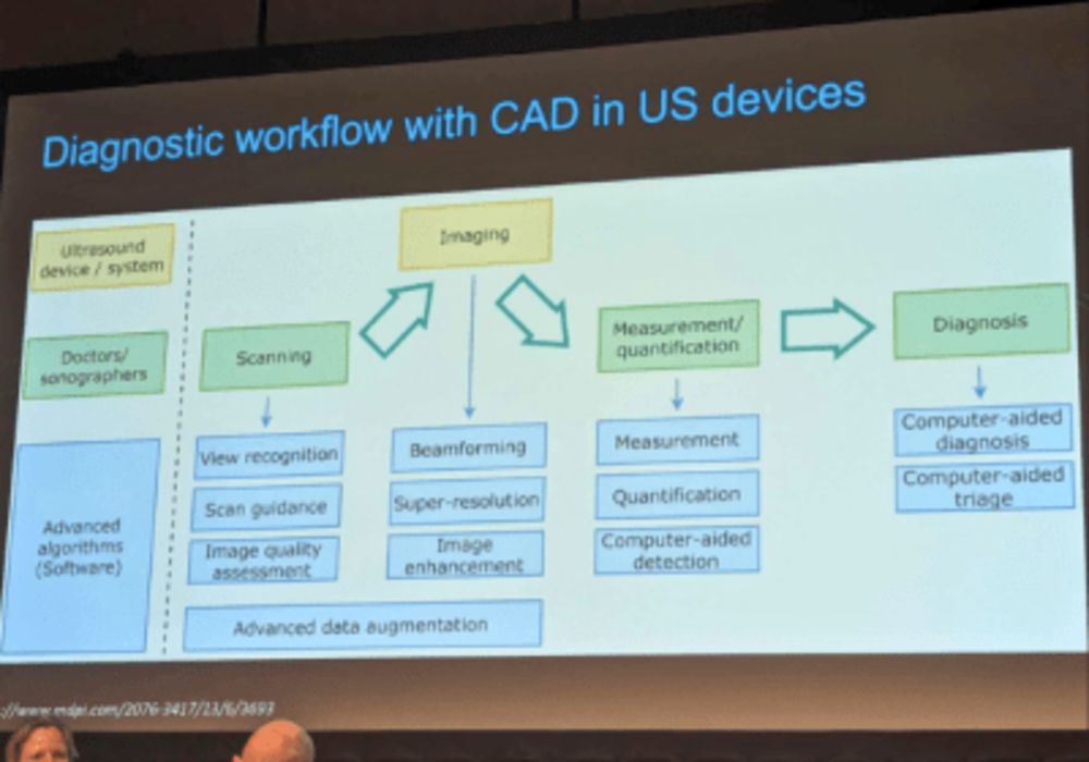
Al software needs to fit into the US workflow differently than for example CT scan. Emphasis must be on real-time analysis at the point of US acquisition vs. CT automated reading after examination. Compared to the image acquisition and analysis abilities of a sonography specialist, no known current Al method is generic enough to be applied on a wide range of tasks. For each ultrasound task AI can be applied to answer various questions:
- Classification “what objects are present in this image?”
- Segmentation “where are the organ boundaries?”
- Navigation “how can I acquire the optimal image?”
- Quality assessment “is this image fit for purpose to make a diagnosis?”
- Diagnosis “what is wrong with the imaged object?”
The Deep-Learning revolution in ultrasound imaging is underway, as image information obtained by ultrasound provides the foundation of input data for the development of a machine-learning algorithm. It is difficult to have a well-classified, structured ultrasound image data set, (complex factors), as variables in the ultrasound image acquisition process produce enormous amounts of unintentional image data, making it difficult Al to create a usable machine learning algorithm in practice.
The main areas where Al is being used to facilitate US is standard plane recognition and organ identification, extraction of standard clinical planes from 3D US volumes and scanning guidance of US acquisitions performed by humans or robots. AI-based detection can offer auto and lesion oriented scanning protocol (standard protocol guidance without missing planes, high- efficiency and quality control with tailored workflow), auto TI-RADS analysis with high accuracy (easy and precise with fully automatic lesion detection, trace/measure, annotation, TI-RADS analysis and report) and systematic management and assessment with multi-lesions and multi-planes. Auto TIRADS/ATA, BTA, EU-TIRADS and K-TIRADS standardized classification and analysis of suspicious lesions and a report to reduce time from repetitive tasks with improved diagnostic confidence, inter-reader agreement and a reduced number of unnecessary biopsies. Workflow improvement and assistance makes clinical routine of multiplanar diagnosis more accurate, productive and easier.
Opportunities for Clinical Al Application
There is substantial research regarding ultrasound Al applications in areas such as the thyroid, breast, and prostate - small parts ultrasound. However, despite the high sensitivity of CAD systems, they have failed due to variable diagnostic performance and low specificity. Recent research has demonstrated improved diagnostic performance for breast cancer using quantitative colour Doppler radiomics features. Research has been further expanded to demonstrate the feasibility of neural networks for activity scoring of arthritis with impressive results compared to a human expert.
In a recent study measuring CEUS performance with or without AI, combined diagnosis was significantly higher than that of Al or CEUS alone. Al has a high diagnostic efficiency, which was helpful for radiologists to make rapid assessment. Al combined CEUS can significantly improve the diagnostic sensitivity and NPV, which was beneficial for the early detection of malignant nodules.
AI Potential and Challenges for Ultrasound Diagnostic
Potential of AI-supported diagnostic is vast. Al algorithms can enhance the quality of US images by reducing noise, optimizing contrast, and improving overall image clarity. This helps in better visualization of vascular structures and lesions. Al algorithms can quantify blood flow parameters, such as perfusion rate, blood volume, and transit time, from CEUS data. This quantitative analysis aids in the characterization of lesions and provides valuable information for treatment planning. Al can automatically detect and segment lesions in US images. This is particularly useful in identifying tumors, cysts, or other abnormalities in organs such as the liver, kidney, and spleen. Al systems can continuously monitor CD/CEUS images in real-time, flagging any significant changes or anomalies. This can be crucial for dynamic assessments, such as monitoring blood flow during interventional procedures.
AI can also play a major role in making workflows more efficient and less time-consuming for specialists. Al can generate automated reports summarizing key findings from US exams. This saves time for specialists and ensures consistency in reporting. Al can integrate US data and findings directly into electronic health records, making it easier for healthcare providers to access and review patient information.
However, some challenges remain, especially regarding interpretability. While Al algorithms can make accurate predictions, it can be difficult to understand how they arrive at their conclusions. The inherent limits in distinguishing normal versus abnormal in continuous variable biologic data may be the most significant limitation for Al in imaging. This can make it challenging for clinicians to trust and interpret the results of Al models and can limit the use of Al in clinical decision- making.
It's essential for healthcare professionals to stay updated with the latest Al developments and ensure that Al systems are validated and integrated into clinical workflows in a responsible under and ethical manner.


