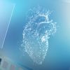


A four dimensional cardiac MRI showing a beating heart frozen in time, has won the British Heart Foundation’s (BHF) annual ‘Reflections of Research’ image competition, revealing stunning images from its research into heart and circulatory disease.
A second image, heart cells of a newborn, was chosen from an online vote on the BHF Facebook page and the winners of the tenth annual competition were formally announced at the British Cardiovascular Society (BCS) Conference in Manchester.
The Reflections of Research Judges’ Winner was “Go with the flow”, by Dr Victoria Stoll, a BHF-funded researcher at the University of Oxford.
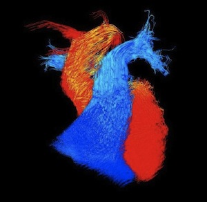
The winning image, using four dimensional cardiac magnetic resonance imaging (MRI), captures the blood flowing within an adult heart frozen in time. Blood flows within the main pumping chambers – the ventricles – of the heart and the vessels leaving the heart. The blue flow is blood that lacks oxygen and is travelling to the lungs. The red flow is blood that has been through the lungs and received oxygen and is now ready to be pumped around the body.
Dr Stoll is using this type of imaging to look at the blood flow in four dimensions within the hearts of people with heart failure, whose hearts are not pumping effectively. She has already found that in people with severe heart failure the blood flows around the heart in a more disordered and disrupted pattern.
The public favourite, “Heart cells of a newborn” by Dr Elisa Avolio, a BHF-funded researcher at the University of Bristol, shows heart cells taken from newborns who received a surgical operation for correction of congenital heart defects shortly after birth. The blue colour shows the shape of the cells, while the magenta shows a structural protein necessary to glue the cells together.
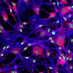
Dr Avolio is using these cells to explore the possibility of treating congenital heart disease, the most common type of birth defect.
“Science relies increasingly on ever more sophisticated imaging techniques to help us to see the cellular and molecular processes that conspire to create disease,” said Professor Peter Weissberg, Medical Director at the BHF and one of this year’s judges.
“Each of these images contains a wealth of information that scientists can use in their fight against cardiovascular disease. So whilst this competition is all about stunning imagery, it’s actually the story that the image tells that matters.”
“The winning image is simply beautiful. It’s both amazingly abstract and instantly recognisable. My 11 month old daughter is fascinated by it, and she is perhaps the best judge of all showing that this image is simple yet also very striking, which is what a good photograph should be,” added wildlife photographer Andrew Rouse one of the four judges.
“Bringing the worlds of art and science together is such a perfect way to explore the wonders of science and the extraordinary insights that we are able to witness through the technology that scientists have access to. Finding an artistic expression can make these concepts accessible to a much wider community,” concluded competition judge and artist, Sophie Layton.
The winners and top ten shortlisted images are available at