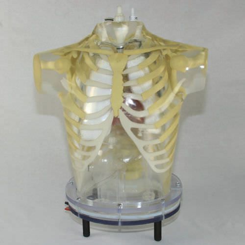
The thorax is molded of polyurethane, modified for tissue-equivalence, with a mass density of 1.10 g/cc. The narrow beam linear attenuation coefficient measured at 140 keV (Tc-99m) is 0.160 cm -1. The skeleton, embedded in the soft tissue, extends from the suprasternal notch down to L2. The RSD materials closely meet the standards of the International Commission on Radiation Units and Measurement (ICRU) Report No. 44 (Tissue Substitutes in Radiation Dosimetry and Measurement, 1989) for both the cortical and spongiosa components of the human skeleton. The mass densities are 1.88 g/cc for cortical bone and 1.16 g/cc for spongiosa. The narrow beam linear attenuation coefficient for the cortical component, measured at 140 keV, is 0.280 cm-1. The volume of the thoracic cavity, when all organs (heart, lungs, and liver) are inserted, is about 8,200 ml. It is filled from the top, with either an inert or a radioactive solution, to simulate the thoracic background. A valve is installed at the base for conveniently draining the phantom. The residue on the walls of the cavity can be easily flushed with the fittings provided at the top of the phantom. A second, smaller fitting is also provided as an air-bleed during filling.
a:2:{i:0;a:2:{s:4:"name";s:20:"Type of calibration:";s:3:"val";s:19:"for nuclear imaging";}i:1;a:2:{s:4:"name";s:17:"Area of the body:";s:3:"val";s:5:"torso";}}


