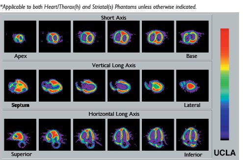
An accurately anatomic heart model is based on vacuum-formed shells. It was designed using high resolution, contrast-enhanced, ultrafast CT data from a normal patient, slightly modified to facilitate its use. The left and right chambers are connected at the atrium region to make a single compartment which can be filled and flushed independently using two ports labeled HC (heart chambers). The right ventricle is slightly modified to allow air to escape during filling. The myocardial wall (MW) has two ports, flushing and independent filling. The volume of the heart chambers is 284 ml, while the volume of the myocardial wall is 238 ml, without inserted defects. The standard model includes three defects with volumes of 8.9, 13.5, and 41.7 ml, respectively. Each of the defects can be filled separately. Defects of different dimensions can be ordered at no added cost. A disassembled heart is sent on request, so that dimensions of a special set can be established. Note that different defects cannot be retrofitted in the assembled heart.
a:2:{i:0;a:2:{s:4:"name";s:17:"Area of the body:";s:3:"val";s:12:"heart muscle";}i:1;a:2:{s:4:"name";s:20:"Type of calibration:";s:3:"val";s:19:"for nuclear imaging";}}


