


A comprehensive understanding of the structurally normal and diseased human heart is crucial for improving the diagnosis and treatment of cardiovascular diseases. Traditional imaging methods like CT scans and MRIs have limitations in resolution and the need for contrast agents. However, recent advancements in synchrotron x-ray phase-contrast imaging, specifically hierarchical phase-contrast tomography (HiP-CT), offer unprecedented insights into cardiac microanatomy. This article explores the capabilities of HiP-CT in imaging intact adult human hearts at cellular resolution, highlighting its potential to revolutionise cardiovascular diagnostics and research.
Advancements in X-ray Phase-Contrast Imaging
Unlike conventional X-ray techniques, phase-contrast imaging exploits the phase shift of X-rays passing through tissues, resulting in images with significantly higher contrast and resolution. This method is particularly advantageous for visualising soft tissues and fine structures, such as those in the heart, without the need for exogenous contrast agents. HiP-CT, a cutting-edge development in phase-contrast imaging, uses the brilliance of fourth-generation synchrotron sources to achieve high spatial resolution. This technique has overcome previous limitations, such as field-of-view and resolution constraints, enabling the imaging of entire adult human organs with cellular detail.
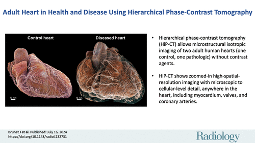
Image Credit: RSNA Radiology
HiP-CT: Bridging Macro- and Microanatomy
HiP-CT represents a breakthrough in cardiac imaging, allowing for the non-destructive, high-resolution examination of the heart's macro- to microanatomy. In a recent exploratory study, HiP-CT was used to image two hearts: one from a 63-year-old male without significant cardiovascular disease and another from an 87-year-old female with multiple cardiac comorbidities. The hierarchical approach began with imaging the entire heart at an isotropic voxel size of approximately 20 µm, followed by local "zoom" scans in regions of interest with voxel sizes down to 2.3 µm. This multiscale imaging provided detailed, comprehensive views of myocardial architecture, cardiac conduction systems, and vascular structures, demonstrating the feasibility of quantitative measurements relevant to clinical cardiology.
Quantitative Analysis and Clinical Relevance
The study demonstrated the potential of HiP-CT for quantitative analysis of cardiac structures, providing a level of detail and accuracy previously unattainable. For instance, the atrial walls' thickness and myocyte aggregates' orientation were measured, providing insights into myocardial architecture and its implications for diseases like atrial fibrillation. Additionally, HiP-CT allowed for the visualisation of mitral annular disjunction, a controversial anatomical feature associated with mitral valve disease. These quantitative capabilities underscore the clinical relevance of HiP-CT in understanding and diagnosing cardiac conditions, instilling confidence in its potential as a diagnostic tool.
HiP-CT is poised to revolutionise cardiac imaging by bridging the gap between whole-organ and cellular scales. Its ability to provide high-resolution, three-dimensional images of the heart without the need for invasive procedures or contrast agents offers new opportunities for cardiovascular research and diagnostics. Despite some limitations, such as the need for specialised equipment and ex vivo imaging constraints, HiP-CT represents a promising tool for advancing our understanding of heart anatomy and disease. Future research and broader access to this technology could pave the way for innovative diagnostic and treatment strategies in cardiovascular medicine.
Source: RSNA Radiology
Image Credit: iStock