


Congenital cervical malformations (CCMs) are rare and complex deformities of the female lower genital tract. These anomalies arise due to the improper fusion of the Müllerian ducts or failure of subsequent canalisation during embryonic development. The prevalence of CCMs is relatively low, ranging from 1 to 1.25 cases per 100,000 individuals. Congenital vaginal agenesis is a common coexisting condition in approximately half of these cases. Given the potential for significant reproductive complications, early diagnosis and appropriate management are crucial. A recent article published in Insights into Imaging aims to provide an in-depth exploration of CCMs, including their diagnosis, classification, and treatment options.
Diagnosis of Congenital Cervical Malformations
CCMs often present in pubertal young women with symptoms such as primary amenorrhea, cyclical abdominal pain, or a pelvic mass. Delayed diagnosis can lead to severe complications, including endometriosis. Magnetic resonance imaging (MRI) is a non-invasive diagnostic tool that offers high soft tissue contrast and multiplanar imaging capabilities, making it essential for early and accurate diagnosis of CCMs. MRI can reveal detailed anatomical features that are crucial for identifying the specific type of cervical malformation.
The diagnostic process involves a thorough clinical evaluation and imaging studies. MRI is particularly valuable as it provides high-quality anatomical information, aiding in differentiating various types of CCMs. Despite its advantages, MRI-based classifications often require confirmation through postsurgical evaluation due to the complexity of the malformations. This underscores the need for a comprehensive and practically employable classification system to guide clinical management effectively.
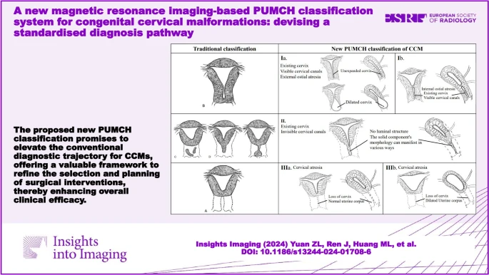
Image Source: Insights into Imaging
Classification Systems and Their Clinical Utility
Various classification systems have been developed to categorise CCMs based on anatomical and imaging features. The European Society of Human Reproduction and Embryology and the American Society for Reproductive Medicine classify CCMs under different categories, highlighting the lack of a universally accepted system. The most widely accepted classification is by Rock et al., based on a study of 30 patients. However, this system primarily relies on anatomical structures identified post-surgery, posing challenges for preoperative diagnosis using imaging alone.
A new classification system proposed by Peking Union Medical College Hospital (PUMCH) aims to address these limitations by integrating MRI features. This system categorises CCMs into three types: Type I (cervix with visible cervical canal), Type II (cervix with invisible cervical canals), and Type III (cervical aplasia). This classification not only aids in the standardisation of diagnosis but also helps in planning appropriate surgical interventions. For instance, patients with Type I CCM may benefit from conservative surgeries, while those with Type II and III may require more extensive reconstructive procedures.
Treatment Options and Outcomes
Historically, hysterectomy has been the treatment of choice for CCMs. However, advances in surgical techniques have introduced conservative options such as canalisation and cervical reconstruction. The choice of treatment depends on the type of CCM and the presence of the vagina. While conservative surgeries aim to restore normal uterovaginal anatomy, they carry risks of complications and failure. Therefore, the selection of the appropriate surgical approach is critical to prevent severe complications that could impair reproductive potential.
The PUMCH classification can guide the choice of treatment. For instance, Type I CCMs, which retain some cervical functionality, may respond well to uterine vaginal penetration or cervicoplasty. In contrast, Type II and III CCMs, characterised by more severe structural anomalies, might necessitate more complex reconstructive surgeries or even hysterectomy in cases where conservative methods fail. The success of these treatments is contingent upon early and accurate diagnosis, underscoring the importance of utilising advanced imaging techniques and a robust classification system.
Congenital cervical malformations, though rare, pose significant challenges in terms of diagnosis and treatment. The early detection of these anomalies is crucial to prevent long-term reproductive complications and improve patient outcomes. MRI plays a pivotal role in diagnosing CCMs, providing detailed anatomical information that is essential for effective clinical management. The development of a new classification system by PUMCH offers a promising tool for standardising the diagnosis and treatment of CCMs, potentially leading to better surgical outcomes and improved quality of life for affected individuals. Future research should focus on refining these classification systems and expanding the cohort of patients to enhance the accuracy and applicability of these diagnostic tools.
Source: Insights into Imaging
Title Image: iStock