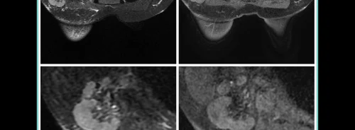
Contrast-enhanced mammography (CEM) is a promising breast imaging method utilising iodinated contrast material and low- and high-energy images to highlight hypervascular lesions. Recent research indicates high diagnostic performance in breast cancer detection, especially when interpreting both types of images together, with sensitivity and specificity values of 95% and 81%, respectively. It serves as an effective alternative to MRI for patients with contraindications to magnetic fields, offering faster and less expensive imaging with higher positive predictive value and fewer false positives. CEM accurately resolves equivocal findings from conventional imaging, preoperative staging, and monitoring response to neoadjuvant therapy. It shows advantages in cost, availability, and reducing false positives compared to MRI in preoperative staging. Integrating CEM with contrast-enhanced computed tomography (CE-CT) for breast cancer staging could optimise the process without additional contrast material, offering potential benefits in evaluating disease extent. Preliminary studies suggest that performing CEM immediately after CE-CT is feasible and does not affect breast lesion detection. Similarly, the aim of a recent study published in European Radiology Experimental was to evaluate the diagnostic potential of CEM performed immediately after contrast-enhanced CT for local staging of BC.
Comprehensive Breast Imaging and Diagnosis: A Prospective Study Integrating Various Modalities
Various breast imaging techniques were utilised in a prospective study involving 50 consecutive patients aged 55.9 ± 9.4 years, referred to the hospital's Breast Unit between January 2020 and March 2022. All patients underwent mammography with digital breast tomosynthesis, breast ultrasonography (US), and core needle sampling to confirm breast cancer (BC) diagnosis. Patients meeting indications for contrast-enhanced computed tomography (CE-CT) based on the American Joint Committee on Cancer (AJCC) manual for BC were enrolled, while those with specific exclusions such as renal failure, contrast agent allergy, pregnancy, prior breast surgery, or implants were excluded. CE-CT was conducted using a 64-slice scanner and iodine-based contrast material, employing a triphasic technique. Following CE-CT, contrast-enhanced mammography (CEM) was performed without additional contrast material injection using automated parameters. Two experienced radiologists evaluated CEM images, particularly focusing on contrast-enhanced images, and classified lesions according to the Breast Imaging Reporting and Data System (BI-RADS) CEM lexicon. Histopathological confirmation was obtained through core needle biopsy, with definitive postoperative histological examination serving as the reference standard for correlating tumour size and extent detected by breast imaging or CEM with histological specimens.
Clinical Insights from Comprehensive Breast Cancer Assessment
Among the 50 patients studied, 60% had unifocal lesions, while 40% had multicentric or multifocal lesions, with 25% of the latter group exhibiting bilateral involvement. A total of 78 malignant lesions were found, predominantly invasive ductal carcinomas (92%) and invasive lobular carcinoma (8%). Neoadjuvant therapy was administered to 24% of patients, with the rest undergoing immediate surgical treatment. Axillary metastatic adenopathies were present in 64% of patients, while internal mammary metastatic adenopathies and distant metastases were found in 22% and 10% of patients, respectively.
Comparing unenhanced breast imaging with contrast-enhanced mammography (CEM), CEM demonstrated a sensitivity of 100%, significantly higher than the 81% sensitivity of unenhanced imaging. Unenhanced imaging missed 15 malignant lesions detected by CEM. However, these additional findings did not alter treatment decisions. Most lesions on CEM were classified as having high conspicuity (74%).
Interobserver agreement between radiologists was high for overall detection of malignant breast lesions (κ value = 0.94), classification of index tumour conspicuity (κ value = 0.95), and evaluation of tumour size and extent (κ value = 0.86). Correlation coefficients for index tumour size and extent with histological specimens were higher for CEM (0.75) compared to unenhanced imaging (0.50).
Contrast-Enhanced Mammography and Integrated Imaging Strategies
The choice of treatment for breast cancer (BC) heavily relies on locoregional and systemic staging, encompassing factors such as tumour size and extent, additional foci of BC, lymph node involvement, and distant metastases. Contrast-enhanced breast MRI is the standard for locoregional staging, offering superior accuracy in tumour size assessment and detection of additional lesions compared to mammography and ultrasound. However, MRI can lead to over- or underestimation, potentially resulting in unnecessary surgeries.
Contrast-enhanced mammography (CEM) has emerged as a promising alternative for BC staging. Studies have shown CEM's efficacy in detecting index tumours and additional lesions, with comparable sensitivity to MRI. While MRI may detect more additional lesions, CEM shows a lower false-positive rate. CEM can influence treatment decisions, particularly in cases requiring surgical management.
In assessing the index tumour size and extent, CEM has shown varied accuracy compared to surgical specimens and MRI. Some studies report CEM to be highly correlated with surgical findings, while others suggest MRI as more accurate. Additionally, CEM has demonstrated potential in preoperative planning, with the ability to detect additional lesions and change surgical management in a subset of patients.
Recent studies have explored the feasibility of performing CEM immediately after contrast-enhanced CT (CE-CT) without additional contrast material injection. This approach offers a potential one-step solution for both locoregional and systemic BC staging. CE-CT provides information on systemic staging, while CEM accurately evaluates local disease extent. Preliminary findings suggest high sensitivity and correlation with histological findings for CEM performed after CE-CT.
While promising, these studies are limited by sample size, logistical constraints, and lack of direct comparison with MRI findings. Further research is needed to validate these findings in larger and more diverse patient populations and to explore the potential role of CE-CT in systemic BC staging. Nonetheless, the integration of CE-CT and CEM could offer an efficient and cost-effective approach to BC staging, warranting further investigation.
Source: European Radiology Experimental
Title Image: iStock


