Accurate, noninvasive assessment of liver fat is key to managing metabolic dysfunction-associated steatotic liver disease. A large prospective project across tertiary centres assessed ultrasound-derived fat fraction as a practical alternative to biopsy and advanced magnetic resonance methods. Adults with suspected disease underwent standardised ultrasound first, followed shortly by a reference test. The work focused on agreement with reference standards, day-to-day consistency and a simple way to turn continuous values into clear decisions. It also explored how a brief sequential pathway using a clinical index could minimise indeterminate results in people with higher body mass index. The findings point to a fast test that fits routine workflows while supporting confident rule-out and rule-in decisions for different levels of steatosis.
Standardised Test with Broad External Comparison
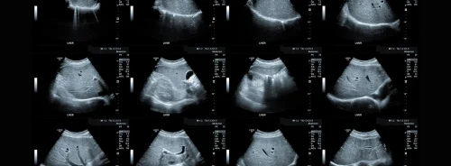
Seven hundred ninety participants were enrolled across 14 hospitals and randomly assigned to derivation and validation cohorts. Each person had ultrasound-derived fat fraction (UDFF) measured using a consistent protocol: a defined region in the right anterior lobe, breath held in neutral and the median of five acquisitions analysed. Examinations took only three minutes, supporting real-world hospital workflow. Reference standards included MRI proton density fat fraction (MRI-PDFF), liver histopathology and proton spectroscopy, enabling comparison against both imaging and tissue-based benchmarks within a short interval after ultrasound.
UDFF aligned closely with each reference method. Agreement with MRI-PDFF was strong, and UDFF values rose stepwise with histological severity from no steatosis to advanced grades. Reproducibility was excellent, with tightly matched measurements on repeat testing and strong alignment between readers with different levels of experience. Against clinical indices used in primary or metabolic care, UDFF showed higher diagnostic performance for all binary tasks evaluated, reinforcing its value as a frontline imaging measure rather than a secondary risk score.
Must Read: Evaluating MRI and Ultrasound for Grading Hepatic Steatosis
Clear Cutoffs and a Manageable Grey Zone
To support decision-making, the analysis derived paired cutoffs for each steatosis band: a lower threshold to rule out disease with high sensitivity and an upper threshold to rule in with high specificity. For example, for any steatosis (S1–S3), 6% served as a rule-out point and 10% as a rule-in point. Similar paired thresholds were set for moderate and severe steatosis. This approach translates a continuous UDFF measurement into immediate next steps: reassurance when below the lower limit, confirmation when above the higher limit and targeted follow-up only when results sit between the two.
The grey zone between thresholds was kept to a minority of cases in both derivation and validation cohorts. Even within this indeterminate band, UDFF still narrowed uncertainty by identifying a much smaller group that actually needed additional triage. This framing avoids over-interpretation of borderline values while giving clinicians a consistent way to apply a single ultrasound measurement across varied settings, including hepatology clinics, metabolic services and radiology departments. It also supports continuity across pathways, since the same paired-threshold logic applies to different severity bands without constant recalibration.
BMI-Aware Pathway Reduces Indeterminate Results
Body mass index (BMI) influenced the dispersion of measurements, with slightly tighter agreement against MRI-PDFF at lower BMI and a somewhat wider indeterminate range at higher BMI. To address this, the investigators tested a simple sequential strategy in people with BMI at or above 23 kg/m². UDFF was applied first using the paired thresholds for any steatosis. For those who remained in the grey zone, the Hepatic Steatosis Index (HSI) was applied at its higher cutoff. This two-step pathway reclassified a substantial share of indeterminate cases as steatosis and reduced the overall indeterminate rate from 18.0% to 7.6% in the higher-BMI subgroup.
In people with BMI below 23 kg/m², UDFF alone already provided strong rule-out and rule-in performance using the same paired thresholds, so sequential testing offered less additional value. Importantly, the sequential pathway did not change the thresholds themselves or the way UDFF was acquired, it simply added a quick, widely available clinical index for those most likely to sit between cutoffs. The result is a practical template that services can adopt without new infrastructure, extending the reach of ultrasound while avoiding unnecessary referrals or invasive tests.
Across multiple centres, ultrasound-derived fat fraction delivered accurate, consistent and time-efficient assessment of hepatic steatosis against imaging and histological reference standards. Paired thresholds turned a single quantitative measurement into clear rule-out and rule-in decisions while keeping the indeterminate zone small. In people with higher BMI, a brief sequential step with the Hepatic Steatosis Index further reduced uncertainty and preserved a high confirmation rate among those reclassified. The approach supports routine integration in radiology and hepatology workflows, offering a practical route to noninvasive grading and risk stratification. Broader external validation and future outcome studies would help confirm generalisability across populations and care settings.
Source: Insights into Imaging
Image Credit: iStock


