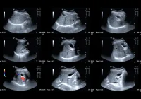
Contrast-enhanced ultrasound (CEUS) has grown from a narrowly licensed tool into a technique with much wider clinical relevance. Initially approved only for focal liver lesions, breast, peripheral arterial system and cardiac imaging, its use has steadily expanded over the past two decades. Clinicians increasingly apply CEUS to a variety of organs and conditions, recognising its advantages over computed tomography (CT) and magnetic resonance (MR) in terms of safety, cost and patient acceptability. CEUS uses microbubble contrast agents that are strictly intravascular, non-nephrotoxic and do not cross the placental barrier, making it especially valuable in vulnerable populations. Despite mounting evidence, controversies persist regarding its application in pregnancy, paediatrics, abdominal trauma, renal cyst evaluation and follow-up after endovascular aortic repair (EVAR). These areas highlight both the promise and the challenges of integrating CEUS more widely into diagnostic pathways.
CEUS in Pregnancy and Paediatric Imaging
In pregnancy, ultrasound remains the frontline imaging method due to its safety and accessibility. While CT and MR are used when more detailed evaluation is necessary, both carry risks: CT involves ionising radiation and MR uses gadolinium-based agents, which raise concerns over foetal safety and environmental contamination. CEUS presents an appealing alternative as its microbubbles remain within the maternal vasculature and do not enter the foetal circulation. Reports describe successful application in conditions such as caesarean scar pregnancy, invasive placenta percreta, uteroplacental blood flow assessment and hepatic lesions during pregnancy. However, major societies have not formally approved its use in obstetrics, citing the limited body of prospective data. For now, CEUS in pregnancy is reserved for carefully considered cases where its diagnostic benefits outweigh uncertainty about long-term safety.
Paediatric imaging illustrates another strong case for CEUS expansion. Children benefit from imaging techniques that avoid radiation and sedation, both of which are common with CT and MR. CEUS offers real-time visualisation of vascularity in solid organs and allows parents to remain present during the examination, making it a more acceptable experience. Evidence supports its use in focal liver lesion characterisation, leading to regulatory approval in the United States for this indication in 2016. Since then, clinical practice has broadened to include trauma assessment, oncology and interventional radiology. Promising results have also been reported for vesicoureteral reflux using CEUS voiding urosonography, which provides a radiation-free alternative to fluoroscopic cystourethrography. Concerns remain about a slightly higher rate of hypersensitivity reactions in children compared with adults, but overall safety profiles are favourable. Ethical and regulatory challenges persist, as most uses remain off-label, but growing evidence is driving wider acceptance.
Trauma and Renal Cyst Management
Blunt abdominal trauma represents one of the most compelling scenarios for CEUS. Standard diagnostic pathways emphasise CT and focused abdominal sonography for trauma, yet CEUS demonstrates significantly higher sensitivity and specificity than conventional ultrasound for detecting organ injuries. It enables assessment of hepatic, splenic, renal, adrenal and pancreatic trauma, correlating closely with CT findings. Its portability allows bedside use and repeated assessments during follow-up, making it especially valuable in emergency settings and resource-limited environments. CEUS is particularly advantageous in paediatric trauma follow-up, where it detects complications such as pseudoaneurysms without exposing children to radiation. Limitations include reduced performance in multi-organ injuries, difficulty with bowel or diaphragmatic lesions, and dependence on operator skill. Regulatory barriers also constrain its integration into major trauma guidelines despite clear clinical value.
Must Read: Improving Liver Lesion Diagnosis with Contrast-Enhanced US
Renal cysts provide another area where CEUS has proven useful. Cystic renal lesions are common, particularly in elderly patients, and their classification determines management. The Bosniak system, traditionally based on contrast-enhanced CT, has guided risk stratification for decades but carries limitations related to radiation exposure, interobserver variability and inadequate characterisation of atypical lesions. CEUS offers superior resolution for evaluating septations, wall thickening and vascularity, features that are key indicators of malignancy. Meta-analyses confirm that CEUS achieves high sensitivity and specificity in differentiating benign from malignant cysts. The 2019 update of the Bosniak classification and subsequent European proposals have incorporated CEUS, showing good reproducibility even among less experienced observers when structured reporting is used. CEUS avoids nephrotoxic agents, can be performed without renal function tests and is more cost-effective than repeated CT or MR follow-up. Challenges remain in patients with unfavourable body habitus or poor acoustic windows, but where feasible CEUS offers immediate and accurate lesion characterisation.
Surveillance After Endovascular Aortic Repair
Long-term surveillance following EVAR for abdominal aortic aneurysm traditionally relies on CT to monitor complications such as endoleaks, graft migration and sac enlargement. Lifelong CT carries cumulative risks of radiation and repeated exposure to iodinated contrast agents, particularly problematic in elderly patients with impaired renal function. CEUS offers a safer alternative, avoiding radiation and nephrotoxicity while delivering high diagnostic accuracy. Studies show CEUS matches CT in detecting endoleaks and may outperform it in identifying late complications such as endotension. Its real-time capabilities and lower cost further enhance its value.
Despite these advantages, CEUS effectiveness can be reduced in patients with high body mass index or excessive bowel gas, and comprehensive visualisation of the entire graft is sometimes difficult. As with other applications, interpretation is operator-dependent and requires specific training. Integration of fusion imaging, which combines CEUS with CT data, further refines anatomical localisation of leaks and supports treatment planning. Wider training programmes and structured protocols could embed CEUS into routine EVAR follow-up, reducing reliance on CT and improving patient safety over the long term.
The evidence for CEUS across pregnancy, paediatrics, trauma, renal cyst evaluation and EVAR follow-up demonstrates its versatility and clinical value. It provides accurate imaging without ionising radiation or nephrotoxic contrast, is well tolerated by patients and can reduce downstream healthcare costs by limiting reliance on CT and MR. Remaining barriers include regulatory approval, limited prospective data in some fields, and the need for specialist training. Nevertheless, CEUS has already established itself as a safe and effective tool beyond its licensed uses. Wider adoption into guidelines and practice could improve diagnostic pathways, protect vulnerable patients from unnecessary risks and support more cost-effective care.
Source: Insights into Imaging
Image Credit: iStock


