


3D ultrasound imaging has been used for several years especially in high-risk Obstetrics. Perinatologists and Maternal Fetal Medicine specialists were among the first to transition to 3D ultrasound and make it a part of their routine exams.
However, the use of 3D ultrasound in the Obstetric office environment has been slower to gain acceptance. The primary reason is the cost of an ultrasound system with the 3D feature. Yet another and more specific reason is the time it takes to learn how to transition from 2D imaging to 3D and 4D imaging.
In 3D ultrasound today, the GE Voluson series has been the market leader. The 3D rendering quality of these systems is excellent which is why it is the choice among most high-risk OB physicians. Even with this excellent rendering quality, for a user new to 3D/4D imaging, these systems are expensive and not that easy to use.
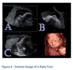
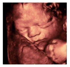
What makes the 2D to 3D transition easier is the user-interface of the ultrasound system. Even before a complete understanding of 3D is known, a user can acquire a baby face routinely which is the most desired image for expecting parents. Acquiring the baby face is the first step to understanding the benefits of 3D ultrasound imaging.
A new system from Mindray Medical, the DC-8, is the solution to the issue of cost and the ease of transitioning from 2D to 3D ultrasound.
From inception, this system was designed for ease of use especially with 3D/4D volume imaging and priced for the OB office market.
What makes the DC-8 easier to use, is that it was designed by clinical users for clinical users. From the Control Panel to the Touch Panel, ergonomics and user ease were considered.
This is especially realized with the 3D/4D interface design.
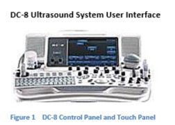
At first glance, the overall system looks like a more advanced ultrasound system. Unlike many less expensive systems, ergonomics and user comfort while reducing scan time by reducing keystrokes was considered in the planning phase.
Advanced features such as 3D/4D Volume Imaging were carefully designed to support the new 3D user and accelerate their learning.
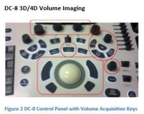
Like most ultrasound systems, controls radiate from a centered Home Base consisting of the Trackball and the buttons that control the functions assigned to the Trackball.
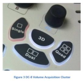
What is unique on this Control Panel is the Volume Acquisition Cluster just above and right of Home Base. Here the 4D Knob and independent 3D Button are surrounded by three Display Format Keys, Single, Dual, and Quad.
Having independent 3D and 4D hard keys is common to more advanced ultrasound systems but having the volume Display Formats around these keys is not. During 4D acquisition or post 3D acquisition, this feature uses a Display Format change more than most other features. The basic reason is so the parents can see the surface rendered baby face in the Single Display Format, however, the imager most often needs to see the A Plane and in many cases the B Plane to acquire the perfect baby face image.
Other quick change functions have also been designed in to make the acquisition of a surface rendered baby face easy and fast for any user. To understand these improvements, a clear understanding of how it is done today is necessary.
4D Volume Acquisition
When acquiring a 4D volume, the user initiates 4D and the first screen is the Set Up. To Set Up for the volume acquisition, simply place the Region of Interest over the anatomy to be acquired. This will most likely be the fetal face. Once the ROI is placed, simply pushing the Update Key will start the acquisition. The user moves the transducer slowly to keep the baby’s face in view and the system surface renders the face. However, 4D resolution is not as good as 3D resolution and the rule of thumb is that if the baby is not moving, a 3D acquisition is better. During a 4D acquisition, the moments a baby stops moving are often times not many and not for long. With almost every ultrasound system, the transition from 4D to 3D, or 3D to 4D, requires the user go through another Set Up. This transition means returning to the Set Up screen, resizing and re-positioning the ROI over the anatomy of interest, then pushing the Update Key to initiate the new 3D acquisition. Moving through another Set Up screen takes valuable seconds during which the fetus will move thus requiring the user to start over again in 4D.
The DC-8 allows an immediate transition between 3D and 4D or 4D and 3D without returning to the Set Up screen. Since the user is already in position during the 4D acquisition, if the baby stops moving, there isn’t any reason to go to another screen to Set Up for the acquisition. Just push the 3D Key and from the position of the 4D acquisition, a higher resolution 3D image is acquired. The time savings is a plus and the fast transition usually means the acquisition is finished by the time the baby moves again.
This is a unique and time-saving transition that will reduce user frustration while increasing the ability to acquire higher resolution 3D images.
Another unique design concept was making the A Plane or the Acquisition View always active after Freeze. Again, to better understand this advantage, an understanding of how it is done currently is needed.
When acquiring a baby face in 4D, the user watches the A Plane. This is called the Acquisition View because it is the scanning view. Scanning while looking at any other plane would be very difficult.
Putting the top line of the graphic, the Cut Plane of the Volume of Interest (VOI), in fluid above the fetal face is the proper placement of the graphic. Keeping the face in the VOI during the acquisition usually results in a very good surface rendering of the face.
Occasionally, the surface rendered face is still obscured and not seen well, and a glance at the B Plane will show the reason. A slight rotational adjustment will likely be necessary in the B Plane. Selecting the B Plane on the Touch Panel to make the rotational adjustment is necessary. This is how the user tells the system which plane they want to adjust. On all systems, the user must make the plane of interest active to make an X, Y, or Z rotational adjustment. Once this change is made, the surface rendered face is now seen more clearly. The image is frozen and a Single Display of the baby face is saved.
Since the B Plane was the last plane chosen, when resuming 4D acquisition, the B Plane would stay active. This is what typically happens on all systems during volume acquisition.
Since the most common rotational adjustment is a Z rotation of the A Plane during acquisition, keeping the B Plane active when resuming scanning is very frustrating. The user is now in live acquisition and begins to adjust the A Plane but that plane is not active. This results in an adjustment of the A Plane that was not desired and this causes frustration on the part of the user.
Therefore, another unique design concept on the DC-8 is to prevent this from happening by always making the A Plane active after Freeze even if another plane has been chosen. This design feature will not only reduce frustration but it makes sense to a clinical user. The user can only scan from the A Plane.
These two design improvements alone support the clinical user during 3D/4D volume acquisition and are not found on more advanced ultrasound systems.
Additional improvements throughout the 3D/4D feature also support the learning of a new user and help to make 3D imaging a routine part of the OB scan for the normal pregnancy.
Benefits of 3D/4D Ultrasound
Transitioning from 2D to 3D/ 4D imaging, in the past, has not been easy due to system cost and additional learning time but the benefits are now within reach with a system designed for the OB/Gyn office. The Mindray DC-8 ultrasound system meets the requirements for this market.
Usually the first step is the acquisition of the baby’s face.
Even though this rendering may not be clinically necessary, it does bring joy to the expectant parents. Many mothers, who are waiting later in life to get pregnant, also feel a sense of comfort and peace when they see the surface rendering of their baby’s face. Presenting a few of these types of images can strengthen the physician/patient relationship and demonstrate that the practice is very current in accepting new technologies.
Mastering the technique just for this acquisition also creates a foundation of understanding of how to clinically use 3D ultrasound.
Once 3D ultrasound is learned, using it can also reduce scanning time since all of the anatomic information can be acquired in one volume dataset. For example, acquiring a 3D volume of the 4-chamber fetal heart and using one of the spin techniques developed by high-risk OB specialists can demonstrate both the LVOT and RVOT in one image and rule out Transposition of the Great Vessels.
The benefits of 3D ultrasound in gynecology are also important. Even without a special transducer that does automatic 3D acquisition, if the Software is in the system, a freehand acquisition of the uterus can be done. The coronal view of the uterus can provide more accurate information about the shape of the uterus, IUD placement, and position of uterine fibroids. The additional clinical information seen in one sagittal uterine acquisition can save time instead of acquiring a series of 2D images and also add more clinical information.
Summary
Transitioning from 2D to 3D/4D ultrasound can be a challenge but, in Obstetrics and Gynecology, 3D is here to stay.
If the choice is to take on this technology, as a new user, choosing a product that will support the learning through a comprehensive system interface designed with the clinical user in mind is best.
The Mindray DC-8 3D/4D user interface is designed for ease which a new or experienced user will appreciate. Even if the application is presenting the baby’s face in a way everyone can feel the joy, it is worth the challenge. Learning to apply 3D technology will enhance a practice and extend diagnostic confidence.
Think of the benefits of adding 3D imaging to your clinical practice. The transition from 2D to 3D/4D can be an easier choice than you think.
Latest Articles
ultrasound, 2D, 3D, imaging
3D ultrasound imaging has been used for several years especially in high-risk Obstetrics. Perinatologists and Maternal Fetal Medicine specialists were among the first to transition to 3D ultrasound and make it a part of their routine exams.
