The scope of ultrasound-guided regional anaesthesia has grown significantly in recent years, and is now a key technique for pain control. Dr. Jens Döffert, Consultant Anaesthetist at Kreisklinikum Calw in Germany, discusses the advances in regional anaesthesia, as described in his book Sonography in anaesthesia: interventional procedures in adults and children(second edition), written in association with his colleague Dr. Ralf Hillmann from Stuttgart.
Dr. Döffert first used ultrasound-guided pain relief in his early days as a senior physician in a spinal centre in Langensteinbach. In 2006, together with Dr. Ralf Hillmann, a colleague from Stuttgart, he was one of the first anaesthetists to qualify as a trainer – now called instructors – for the German Association of Ultrasound in Medicine (DEGUM). In the same year, the two specialists wrote a textbook about ultrasound-guided regional anaesthesia in adults and children.
Dr. Döffert has continued to focus on regional anaesthesia throughout his career, from several smaller hospitals to a maximum care hospital, as the chief physician in a dedicated orthopaedic unit in Bad Wildbad and, since July 2016, on an even larger scale in Calw. Although a relatively small district general hospital, with just under 200 beds, Calw has a comprehensive orthopaedic service, including specialist spinal surgery. Dr. Döffert works in close association with the spinal surgery team, as he explained: “I mainly deal with orthopaedic and trauma patients – including an upper and lower limb service – and provide pain control for spinal patients. Back pain is a common problem – due to prolapsed discs, narrowing of the spinal canal, or non-specific pain. The demand for ultrasound-guided intervention is rising as patients have become increasingly aware of the risks associated with radiation from multiple Xrays and CT-guided procedures; this is now a major issue for many people. From the comfort point of view as well, patients must lie in a certain position on a hard table for X-rays, often in a cold room, which is even more uncomfortable and painful when you are suffering from back pain. Many people are also apprehensive of lying in a CT ‘tube’. In contrast, ultrasound has none of these disadvantages; I can scan patients in their soft hospital beds, and they can position themselves however they feel most comfortable, as long as I can access the area of interest with my ultrasound transducer. In this way, I can inject into sacroiliac joints, nerve roots and facet joints in the lumbar and cervical areas of the spine, and even perform stellate ganglion blocks. I can also treat problems of the piriform muscle, and perform peripheral nerve blocks where necessary.”
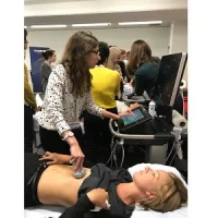
In his day-to-day practice, Dr. Döffertuses a SonoSite S Series™ point-of-care ultrasound system, relying on its excellent image quality to boost presentations and training materials. He added: “I use this machine a lot, and like it very much. It is still perfect, with image quality as good as it was on day one. I am a member of the anaesthesia sector and the interventional sonography working party for DEGUM, and run courses several times a year, using images from this instrument and a SonoSite X-Porte® system for my presentations, as well as for the second edition of our book (published by Elsevier in 2016), the first book to cover ultrasound-guided interventional pain therapy in the German language.”
There have been many developments in ultrasound-guided regional anaesthesia in the past 10 years, most notably the rise in using ultrasound to guide minimally invasive pain control, which is now featured in the second edition. Dr. Döffert continued: “We have added several new access routes, including transversus abdominis plane blocks, and some instances of a change in recommended technique, for example supraclavicular and interscalene access, where we now approach the plexus laterally to avoid anaesthetising the phrenic nerve.”
He concluded: “The beauty of ultrasound guidance for these blocks, and
any pain relief, is that you scan the target area, see where you want to go and
mark it, and the injection itself takes only a minute or two. It is really fast
because the structures are clearly shown, and you are successful in the first
attempt much more frequently than before, which is so important for patients
and anaesthetists alike.”
For more information, please click here.
Source & Image Credit: Sonosite

