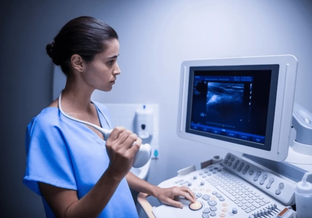
Minimally invasive diagnostic procedures have become increasingly central in gynaecological oncology, particularly ultrasound-guided biopsies. These offer a reliable method to obtain adequate tissue samples for diagnosis, reducing both procedural risk and patient burden. Despite their growing use, there has been a lack of comprehensive guidelines to support clinicians in adopting and refining these practices. To address this gap, the International Society of Ultrasound in Obstetrics and Gynecology (ISUOG) and the European Society of Gynaecological Oncology (ESGO) have collaborated on a robust consensus statement. This document outlines evidence-based standards and recommendations for image-guided biopsies, aiming to unify best practices and improve clinical outcomes.
Defining the Role and Scope of Image-Guided Biopsy
Image-guided biopsy is a cornerstone of personalised care in gynaecological cancer, offering critical support for diagnosis and treatment planning. The procedure is especially valuable in cases involving inoperable tumours, suspected recurrence or disseminated disease. As real-time guidance, particularly via transvaginal or transrectal ultrasound, offers close proximity to pelvic tumours, it enhances diagnostic yield while maintaining a favourable safety profile. These procedures can often be performed in an outpatient setting, expediting treatment initiation and reducing patient stress.
The ISUOG/ESGO consensus confirms that ultrasound should be the first-line imaging modality for guidance due to its accessibility, real-time capabilities and dynamic visualisation. Various approaches—transvaginal, transcervical, transrectal and percutaneous—can be adapted based on lesion location and accessibility. The use of Doppler and contrast-enhanced ultrasound further refines the identification of viable tumour regions, ensuring accurate sampling. Ultrasound-guided biopsy is not only safer and more cost-effective than surgery, but also essential for genomic and histopathological analyses required for personalised therapies. Integration within a multidisciplinary team setting is crucial to ensure optimal procedural planning and interpretation.
Optimising Biopsy Techniques and Diagnostic Yield
The consensus identifies two primary techniques for sampling: core-needle biopsy (CNB) and fine-needle aspiration (FNA). While both have demonstrated acceptable levels of diagnostic adequacy and safety, CNB is generally preferred due to its ability to provide larger, intact tissue samples. These samples are more suitable for immunohistochemical and molecular testing, which are vital for tumour classification and treatment decisions. CNB is especially advantageous when evaluating fibrotic or mesenchymal tumours, where cellular detail and tissue architecture are crucial.
Recommended Read: Non-invasive Imaging Techniques To Evaluate Breast Cancer Characteristics
To maximise diagnostic accuracy, at least two 10-mm long samples using an 18-G needle or larger are recommended, with three cores advised for uterine mesenchymal tumours or suspected lymphomas. Despite the larger needle size, the procedure maintains a low complication rate, with serious adverse events occurring in fewer than 1.5% of cases. Minor complications, such as discomfort or transient bleeding, are usually self-limiting.
In scenarios where ultrasound visualisation is suboptimal—due to obesity, ascites or acoustic shadowing—alternative imaging techniques like CT, MRI or image fusion may be employed. While these methods offer complementary benefits, ultrasound remains the most versatile and accessible choice. The consensus also highlights the value of biopsy in clinical trials and targeted therapy planning, supporting its broader use beyond initial diagnosis.
Guidelines for Indications, Contraindications and Safety Measures
A critical component of the consensus is detailed guidance on clinical indications and patient safety. A biopsy should be considered when the result directly informs treatment, especially in primary inoperable cancers, atypical uterine lesions or suspected recurrences. The document outlines relative contraindications, such as bleeding disorders, anticoagulation therapy and difficult lesion access, which should be managed through careful risk assessment and multidisciplinary consultation.
Procedures are categorised by bleeding risk, with most gynaecological biopsies considered low risk. High-risk procedures include biopsies of hypervascular or intra-abdominal visceral lesions. In patients with elevated bleeding risk, routine laboratory screening—including platelet counts, INR and aPTT—is advised. Specific guidance is provided for managing anticoagulation and antiplatelet therapies before and after biopsy, including the use of bridging therapies where necessary.
The consensus also standardises procedural protocols to ensure consistency and safety. These include steps for patient preparation, equipment sterilisation, anaesthesia use and post-procedure care. Special attention is given to patient comfort, consent and anxiety reduction, which are critical for ensuring cooperation and successful outcomes. Ultrasound-guided biopsy should only be performed by clinicians trained in gynaecological ultrasound and interventional techniques, with minimum level II competency recommended for low-risk cases and level III for high-risk procedures.
The ISUOG/ESGO Consensus Statement offers an essential framework for integrating ultrasound-guided biopsy into routine gynaecological oncology practice. By addressing all stages of the procedure—from clinical indication and technique selection to safety protocols and quality assurance—it provides clinicians with the tools to deliver high-quality, evidence-based care. The emphasis on multidisciplinary collaboration, patient-centred communication and diagnostic precision reflects a broader shift towards personalised, minimally invasive oncology. As ultrasound-guided biopsy continues to evolve, this consensus will play a pivotal role in promoting safe and effective practices across diverse clinical settings.
Source: Ultrasound in Obstetrics & Gynecology
Image Credit: Freepik


