Researchers in
Spain are involved in a European network, headed by CERN, to develop an
endoscopic scanner for early diagnosis of some cancers that have a high
mortality rate, such as pancreatic cancer that has a mortality rate up to 90
percent nowadays.
In spite of the latest advances in cancer detection and diagnosis, some types of cancers are detected in advanced stages due to their morphology and location. Improving an early detection of the different types of cancers is essential to increase the survival rates of this disease.
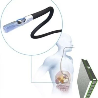
The Endo TOFPET-US project aims to develop a generation of medical scanners specifically designed for the examination of certain organs. This is an international consortium which uses the latest advances on detectors of high energy physics to enhance the quality of nuclear medical images, particularly the images known as Positron Emission Tomography (PET).
Researchers from Biomedical Image Technologies (BIT) at Universidad Politécnica de Madrid (UPM) are involved in the project through the PicoSec educational project and have collaborated in the design and implementation of both the electronics and data acquisition system of the developed sensor. This work was carried out along with the lab of Experimental High Energy Physics and Associated Instrumentation in Portugal (LIP) that gave as a result the spin-off company PETsys Electronics SA.
The latest innovations in detectors for high energy physics carried out in CERN are exceeding the speed limit when detecting elementary particles. PET medical imaging is one application of this technology and consists in a technique that detects pairs of gamma rays emitted indirectly by a positron-emitting and which is used in medical scanners for cancer diagnosis.
As part of the Seventh Framework Programme, the European Union funded EndoTOFPET-US in which various multidisciplinary groups joined forces to develop the first endoscopic PET scanner for specific organs.
The project includes two trends in medical imaging: detectors for examination of certain organs and a multimodal imaging technique, in order to provide data about the cancer that is not available so far. In the case of conventional PET scanners, the patient's body is introduced in a ring of detectors to obtain a cross-section image. Given the new possibilities of miniaturisation, researchers are studying a new asymmetric architecture in which a miniaturised detector is introduced inside the patient's body and placed close to the organ of interest. As a result, this proximity provides higher sensitivity since the patient would receive lower radioactive dose to visualise the lesion without losing the image quality.
In addition to this sensitivity, the new endoscopic detector includes other innovative features. According to UPM researchers: “thanks to its high-speed electronics, this scanner measures the time of flight of photons and this allows researchers a precise identification of the origin point in where particles are concentrated in the tumour mass, filtering the background noise and giving as a result clear images”.
Researchers also add: “the system has a high degree of pixels, providing higher spatial resolution of the image and detecting millimeter lesions”.
The project also includes another trend in biomedical imaging, the multimodal imaging, a way to obtain information by combining diverse techniques and merging the resulting images. Specifically, the scanner combines ultrasounds (US), which give morphological information of the area of interest, with PET, that provides metabolic information to identify cancerous cells.
This innovative scanner requires a high-performance data acquisition system. Thus, UPM researchers in collaboration with PETSys Electronics SA have designed an intelligent, distributed and asymmetric system, which manages a great volume of data due to the large number of channels with different data rates for each type of detector (Endoscopic and abdominal detectors).
Part of the success of this system is based on the decentralisation, since this system moves part of its complexity to the electronics embedded in the detectors. Therefore, the system can implement a multi-level triggering scheme with various stages of filtering data depending on the information available on each stage.
According to UPM researchers: “this new generation of endoscopic scanners will contribute the development of devices that will allow us to visualise cancers in the early stages, and consequently to enhance their prognosis”.


