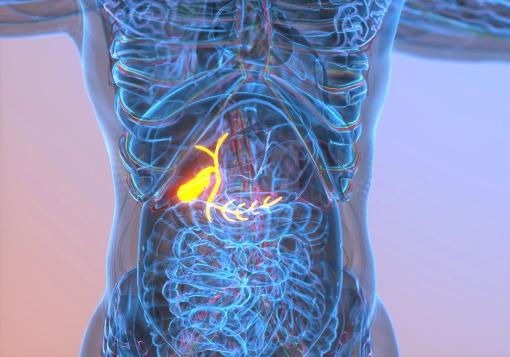
Gallbladder cancer is aggressive with poor prognosis, and curative surgery is often not feasible because most cases are diagnosed at advanced stages. For unresectable or metastatic disease, immunotherapy-based combinations have become first line. Reliable, non-invasive markers that can guide prognosis and treatment selection are therefore a priority. While laboratory and genomic indicators are used, genetic testing is time-consuming and costly, and immune markers such as PD-L1 and CD8+ cell counts show inconsistent prognostic value. Growing attention has turned to tumour-associated tertiary lymphoid structures, which have been linked with outcomes across several solid tumours. An MRI-based radiomics approach now offers a preoperative route to estimate intratumoural tertiary lymphoid structure status in gallbladder cancer and stratify survival after surgery and during immunotherapy.
MRI Radiomics For Preoperative TLS Prediction
Two independent cohorts from separate centres, comprising 129 and 44 patients with gallbladder cancer, were evaluated using preoperative multiparametric liver MRI acquired within one month before surgery. Six sequences were analysed: non-enhanced T1-weighted, T2-weighted, diffusion-weighted, and three contrast-enhanced phases (arterial, portal venous, delayed). Tumours were manually segmented to create volumes of interest. From these datasets, 642 quantitative features per patient were extracted, then reduced through variance and correlation filtering to 78, before multivariate logistic regression selected eight features for a radiomics score. Robust inter-observer agreement supported feature reliability.
Must Read: MRI Radiomics for Liver Tumour Differentiation
Clinico-radiological variables were assessed independently. Three features emerged as predictors of intratumoural tertiary lymphoid structure status: smaller tumour height, absence of liver invasion, and absence of arterial-phase hypo-enhancement. These were incorporated into a clinical model. The radiomics score and the three clinical predictors were then combined into a single model using a weighted formula, creating a combined-score for each patient. Model cut-offs were chosen by the maximum Youden index from receiver operating characteristic analysis and used to classify patients into low- and high-risk groups.
Histopathology provided the reference for intratumoural tertiary lymphoid structures, defined by the presence of secondary lymphoid follicles with germinal centres. In this material, intratumoural tertiary lymphoid structures were confirmed as independently associated with recurrence-free survival, and recurrence was tracked by MRI, CT or PET on a scheduled follow-up pathway. The final follow-up was on 10 June 2023.
Model Development and External Validation
The combined model surpassed either the clinical or radiomics components alone in the training cohort. Areas under the curve were 0.891 for the combined model compared with 0.870 for radiomics and 0.775 for the clinical model. Sensitivity, specificity and accuracy in the training cohort were 0.943, 0.729 and 0.845 for the combined model. These findings were supported in external validation where the combined model achieved an AUC of 0.885, with sensitivity 0.889, specificity 0.824 and accuracy 0.864. Comparative performance was assessed using the DeLong test, and calibration was favourable with non-significant Hosmer–Lemeshow statistics in both cohorts. Decision curve analysis further indicated clinical utility of the combined approach.
Radiomics alone delivered strong discrimination, but integrating clinical context provided incremental gains. The observed pattern suggests that high-dimensional texture and shape information captures intratumoural heterogeneity aligned with immune microenvironments, while tumour height, liver invasion and arterial-phase enhancement characteristics add complementary biological signal. The approach offers a reproducible framework that can be tested across imaging platforms, as demonstrated by external validation.
The analytical pipeline conformed to conventional statistical practices. Univariate screening was followed by multivariate modelling to derive independent predictors. The combined-score drew on coefficients estimated from training data, and risk groups were defined by an empirically determined cut-off. Survival analyses used Kaplan–Meier estimates with log-rank testing. The modelling environment included SPSS, R and Python.
Risk Stratification and Treatment Relevance
Risk groups defined by the combined model separated outcomes meaningfully. In the training cohort, median recurrence-free survival in the low-risk group was 59.6 months compared with 8.7 months in the high-risk group. Six-, 12- and 24-month recurrence-free survival rates were 84.8%, 63.5% and 53.5% in the low-risk group and 53.4%, 40.1% and 36.5% in the high-risk group. In the external validation cohort, median recurrence-free survival was 18.4 months in the low-risk group and 4.2 months in the high-risk group. These gradients indicate that the preoperative imaging signature aligns with clinically relevant trajectories after surgery.
The model also demonstrated relevance in an immunotherapy cohort of patients with advanced gallbladder cancer who had declined or progressed after first-line chemotherapy. In this group, the low-risk category showed a longer median overall survival of 24.7 months versus 10.5 months in the high-risk category. This suggests that the combined MRI-based score may also inform expectations for outcomes in systemic therapy settings, complementing laboratory or genomic markers that are resource intensive or variably predictive.
Intratumoural tertiary lymphoid structures were associated with longer recurrence-free survival on unadjusted analysis and remained independently linked to recurrence-free survival on multivariable modelling. By providing a non-invasive estimate of intratumoural tertiary lymphoid structure status before surgery, the MRI-based approach addresses an important limitation of relying solely on postoperative pathology to identify favourable immune microenvironments.
A combined MRI radiomics and clinico-radiological model estimated intratumoural tertiary lymphoid structure status preoperatively and stratified recurrence-free survival after surgery and overall survival during immunotherapy in gallbladder cancer across two centres. The approach delivered strong discrimination, external validation, sound calibration and decision-analytic support, indicating readiness for further evaluation. Limitations include retrospective design, a small external dataset and manual tumour segmentation, which argue for prospective multicentre studies with larger, balanced cohorts and exploration of automated segmentation. These findings point to a practical imaging-based marker that may support preoperative risk assessment and therapeutic planning without additional invasive testing.
Source: Insights into Imaging
Image Credit: iStock


