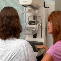
New research shows that adjunct mammography is warranted for evaluation of palpable breast lumps in women age 30 years and older because of the value added to clinical management. In all age cohorts (30–39, 40–49, and 50 years old and older), mammography contributed to delineation of disease extent, detection of incidental malignancies, and confirmation of benign diagnoses, according to the study published in the American Journal of Roentgenology.
Palpable breast lumps are a cause of worry for both patients and their care providers. Although many ultimately have a benign outcome, patients with those lumps have a higher probability of breast cancer than all patients who undergo diagnostic breast imaging. Current guidelines for imaging of patients with a palpable breast lump differ according to patient age. Mammography is the primary imaging modality (followed by ultrasound) for those 40 years and older, and ultrasound is the primary modality for those younger than 30 years.
The superior accuracy of combined mammography and breast ultrasound has been widely published regarding palpable breast lumps. Given recent advances in breast ultrasound, however, concerns related to mammographic radiation, and the decreased mammographic sensitivity posed by dense breasts, it is unclear whether mammography still represents the most appropriate initial study for younger, premenopausal women. The AJR study aimed to determine whether mammography adds clinical value in the diagnostic imaging workup in younger women presenting with palpable breast lumps.
Researchers retrospectively identified the records of all women 30 years old and older, who underwent imaging evaluation with mammography and ultrasound for a palpable lump between 1 January 2009 and 31 December 2010 in a single centre. Benign versus malignant outcomes were determined by pathologic analysis or 24-month imaging or clinical follow-up. The contribution of mammography to final diagnosis was assessed on the basis of objective criteria to determine the clinical impact of mammographic findings.
The study cohort included 861 patients presenting with 935 palpable lumps. Imaging correlates were reported for 568 of 935 (60.7%) lumps, and imaging findings were negative in 367 of 935 (39.3%). Of the 935 palpable lumps, 858 (91.8%) were benign and 77 (8.2%) were malignant. Mammography added clinical value in the evaluation of 27 of 77 (35.0%) malignant lumps by better delineating extent of disease and in the evaluation of 26 of 858 benign lumps (3.0%) by confirming benignity.
Moreover, 52 of 861 (6.0%) patients had incidental findings that led to a recommendation for biopsy. Twenty-nine of the 52 findings were originally seen with mammography and 23 with ultrasound. Mammography also depicted seven incidental malignancies in nonpalpable areas, and ultrasound depicted one incidental malignancy.
"Our results are concordant with literature reports of false-negative rates of combined mammography and ultrasound of palpable breast lumps. Although the false-negative rate is exceptionally low, the presence of malignancy in patients with normal imaging findings at mammography and ultrasound highlights the importance of the clinical breast examination for identifying cases that may warrant further imaging and histologic sampling," the authors wrote. They also noted that mammography alone did not depict any malignancies presenting as palpable abnormalities – this finding was similar to previously published findings.
Owing to the small sample size, the researchers are unable to draw statistically significant conclusions about the 30-39 age group. However, there was a trend towards the contribution of mammography both in identification of malignant disease and confirmation of benign disease. "A larger study focusing on this age group is a worthwhile goal for better understanding the value of mammography to these patients," the researchers added.
Source: American Journal of Roentgenology
Image Credit: Pixabay
Latest Articles
Mammography, palpable breast lump, palpable abnormalities
New research shows that adjunct mammography is warranted for evaluation of palpable breast lumps in women age 30 years and older because of the value added to clinical management. In all age cohorts (30–39, 40–49, and 50 years old and older), mammography