According to researchers, a popular surgery to repair meniscal tears may raise the risk of osteoarthritis and cartilage loss in some patients. The findings, presented at the annual meeting of the Radiological Society of North America (RSNA), suggest that the decision for surgery requires careful consideration in order to prevent accelerated disease onset.
The meniscus is a wedge-shaped piece of cartilage in the knee that serves as a shock absorber between the femur (thigh bone) and tibia (shin bone). Each knee has two menisci that play an important role in joint stability. Meniscal tears are amongst the most common knee injuries, and surgery is often performed to alleviate pain.
"Meniscal surgery is one of the most common orthopaedic procedures performed to alleviate pain and improve joint function," said Frank W. Roemer, MD, from Boston University School of Medicine in Boston and the University of Erlangen-Nuremberg in Erlangen, Germany. "However, increasing evidence is emerging that suggests meniscal surgery may be detrimental to the knee joint."
For this study, Dr. Roemer's team analysed data from the Osteoarthritis Initiative, a large, ongoing observational study of knee osteoarthritis incidence and progression. Patients in the study were on average 60.2 years old and mostly overweight, with a mean body mass index (BMI) of 28.3. About two-thirds of the patients were women.
The investigators reviewed magnetic resonance imaging (MRI) exams of 355 knees that developed osteoarthritis during a five-year period, and a control group that was matched for age, gender, arthritic severity in both knees, and BMI. Of all knees, 31 underwent meniscal surgery during the year prior to the arthritis diagnosis, while 280 knees had signs of meniscal damage on MRI but did not have surgery. Control cases with no meniscal damage were included in the analysis.
Dr. Roemer et al. evaluated the risk of developing arthritis and cartilage loss during the following year for the different groups in the study. "We found that patients without knee osteoarthritis who underwent meniscal surgery had a highly increased risk for developing osteoarthritis and cartilage loss in the following year compared to those that did not have surgery, regardless of presence or absence of a meniscal tear in the year before," Dr. Roemer said, citing these important findings:
An alternative to surgery is conservative management, the research team said. In conservative management, physical therapy is prescribed to help maintain and restore muscle strength and range of motion. Symptoms are usually treated with ice and non-steroidal anti-inflammatory medications.
"The indications for meniscal surgery might need to be discussed more carefully in order to avoid accelerated knee joint degeneration," Dr. Roemer added. Other members of the research team are: Ali Guermazi, MD, PhD; David J. Hunter, MD, PhD; C. Kent Kwoh, MD; Michael Hannon, MA; and Jason Grago, MA.
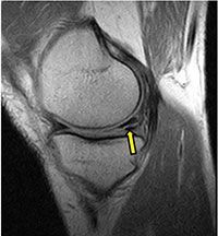
Figure 1A. Examples of meniscal tears and status after surgery. A. Sagittal intermediate-weighted fat-saturated image shows a typical horizontal-oblique meniscal tear of the posterior horn reaching the undersurface of the meniscus (arrows).
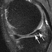
Figure 1B. Examples of meniscal tears and status after surgery. B. Coronal proton density-weighted image shows another example of a tear, which represents a vertical tear of the posterior horn of the medial meniscus (arrow).
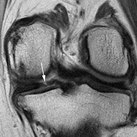
Figure 1C. Examples of meniscal tears and status after surgery. C. Sagittal intermediate-weighted fat-saturated image post surgery shows substance loss of the posterior horn of the medial meniscus (large arrows) and concomitant cartilage loss at the central medial tibia and femur (small arrows).
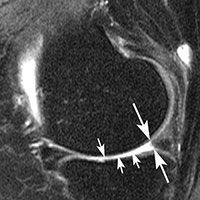
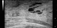
Figure 2. A horizontal tear of the posterior part of the meniscus of the knee.
Source: RSNA
Image Credit: RSNA
The meniscus is a wedge-shaped piece of cartilage in the knee that serves as a shock absorber between the femur (thigh bone) and tibia (shin bone). Each knee has two menisci that play an important role in joint stability. Meniscal tears are amongst the most common knee injuries, and surgery is often performed to alleviate pain.
"Meniscal surgery is one of the most common orthopaedic procedures performed to alleviate pain and improve joint function," said Frank W. Roemer, MD, from Boston University School of Medicine in Boston and the University of Erlangen-Nuremberg in Erlangen, Germany. "However, increasing evidence is emerging that suggests meniscal surgery may be detrimental to the knee joint."
For this study, Dr. Roemer's team analysed data from the Osteoarthritis Initiative, a large, ongoing observational study of knee osteoarthritis incidence and progression. Patients in the study were on average 60.2 years old and mostly overweight, with a mean body mass index (BMI) of 28.3. About two-thirds of the patients were women.
The investigators reviewed magnetic resonance imaging (MRI) exams of 355 knees that developed osteoarthritis during a five-year period, and a control group that was matched for age, gender, arthritic severity in both knees, and BMI. Of all knees, 31 underwent meniscal surgery during the year prior to the arthritis diagnosis, while 280 knees had signs of meniscal damage on MRI but did not have surgery. Control cases with no meniscal damage were included in the analysis.
Dr. Roemer et al. evaluated the risk of developing arthritis and cartilage loss during the following year for the different groups in the study. "We found that patients without knee osteoarthritis who underwent meniscal surgery had a highly increased risk for developing osteoarthritis and cartilage loss in the following year compared to those that did not have surgery, regardless of presence or absence of a meniscal tear in the year before," Dr. Roemer said, citing these important findings:
- All 31 of the knees that underwent meniscal surgery during the prior year developed osteoarthritis, compared with 165 (59 percent) of the knees with meniscal damage that did not have surgery.
- Cartilage loss was much more common amongst knees that had undergone surgery: 80.8 percent of knees with surgery showed cartilage loss, compared with 39.5 percent of knees with meniscal damage and no surgery.
An alternative to surgery is conservative management, the research team said. In conservative management, physical therapy is prescribed to help maintain and restore muscle strength and range of motion. Symptoms are usually treated with ice and non-steroidal anti-inflammatory medications.
"The indications for meniscal surgery might need to be discussed more carefully in order to avoid accelerated knee joint degeneration," Dr. Roemer added. Other members of the research team are: Ali Guermazi, MD, PhD; David J. Hunter, MD, PhD; C. Kent Kwoh, MD; Michael Hannon, MA; and Jason Grago, MA.
Figure 1A. Examples of meniscal tears and status after surgery. A. Sagittal intermediate-weighted fat-saturated image shows a typical horizontal-oblique meniscal tear of the posterior horn reaching the undersurface of the meniscus (arrows).
Figure 1B. Examples of meniscal tears and status after surgery. B. Coronal proton density-weighted image shows another example of a tear, which represents a vertical tear of the posterior horn of the medial meniscus (arrow).
Figure 1C. Examples of meniscal tears and status after surgery. C. Sagittal intermediate-weighted fat-saturated image post surgery shows substance loss of the posterior horn of the medial meniscus (large arrows) and concomitant cartilage loss at the central medial tibia and femur (small arrows).
Figure 2. A horizontal tear of the posterior part of the meniscus of the knee.
Source: RSNA
Image Credit: RSNA
Latest Articles
Arthritis, osteoarthritis, RSNA 2014, #RSNA14, knee surgery, meniscus
According to researchers, a popular surgery to repair meniscal tears may raise the risk of osteoarthritis and cartilage loss in some patients. The findings...