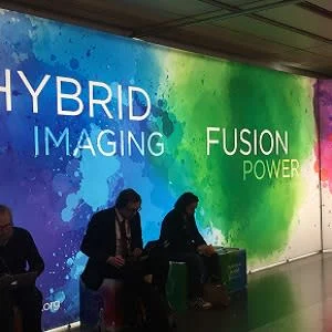
Hybrid imaging and the global trends surrounding the discipline enjoyed special focus at this year’s European Congress of Radiology in Vienna. Hybrid imaging is defined as the fusion of two or more imaging technologies into a single new form of imaging.
The discussion this year concerned this new form of imaging as synergistic and more powerful than the sum of its parts. Despite the fact that certain hybrid imaging modalities may be applied purely to depict anatomy, the most exciting characteristic of hybrid imaging seemed to be its potential to show molecular processes in vivo within their larger anatomic context.
Existing hybrid imaging modalities comprise PET/CT, SPECT/CT, PET/MR, SPECT/MR, ultrasound and MR, ultrasound and CT, MR and CT. The general benefits of hybrid imaging include increased diagnostic accuracy, a further step towards individualised medicine, precise monitoring of interventional procedures and reduced radiation exposure.
You may also like: #ECR2019: AI will be driven by radiologists
A plethora of sessions were dedicated to hybrid imaging at ECR2019 including presentations on cost action initiatives as a platform for image biomarker selection, the role of AI in the introduction of imaging biomarkers, the infrastructure necessary to leverage AI for selecting, validating and managing imaging biomarkers, the clinical impact of hybrid interventional imaging, the optimisation and dose management in hybrid interventional suites and the clinical impact of hybrid interventional imaging.
Latest generation SPECT/CT cameras were presented and how they incorporate multi-detector CT and state-of-the-art gamma camera technology in tandem. These scanners improve the efficacy of a wide variety of nuclear medicine tests by providing more accurate localisation of lesions, exclusion of potentially misleading physiological uptake, characterisation of equivocal or indeterminate activity and detection of additional lesions. In addition, they offer the potential for a more efficient "one-stop-shop" imaging approach. Iterative reconstruction algorithms and faster processing power facilitate radiation dose reduction and increased image resolution. The clinical utility of SPECT/CT is diverse, and a cross-spectrum of applications in musculoskeletal, oncological, cardiological, endocrine, hepatobiliary and GI tract imaging was presented.
There was also an overview of the technological state of the art in PET-MRI, as well as an outlook on emerging new technologies. In this context, new detector set-ups providing higher sensitivity and spatial resolution as well as methods to improve absorption correction and quantification were explained. Furthermore, new PET-insert solutions for Preclinical Research and clinical application were highlighted. An example of the latter, the concept and initial results for a new PET-insert dedicated to human Breast imaging will be presented. In the final part of the talk, the focus was set on the medical applications of PET-MRI. In this context, the discussion considered which applications inevitably demand PET-MRI hybrid imaging and the high potential and clinical value of PET-MRI and its potential future role in patient management.
There is increasing evidence to support a role for metabolism in tumour development; for example, deregulation of cellular energetics is considered to be one of the key hallmarks of cancer. Changes in tumour metabolism over time are now known to be early biomarkers of successful response to chemotherapy and radiotherapy. There are a number of imaging methods that have been used to probe cancer metabolism: the most widely available is 18F-fluorodeoxyglucose (FDG), an analogue of glucose, used in PET. Hyperpolarised carbon-13 MRI (13C-MRI) is an alternative and emerging molecular imaging method for studying cellular metabolism.
This technique allows non-invasive measurements of tissue metabolism in real-time. To date, the most promising probe used in conjunction with hyperpolarised MRI has been 13C-labelled pyruvate: pyruvate is metabolised into lactate in normal tissue in the absence of oxygen, but in tumours, this occurs very rapidly even in the presence of oxygen. Results from many animal models have shown that there is a reduction in the metabolism of pyruvate following successful treatment with chemotherapy. Tumour lactate labelling has also been shown to correlate with the grade of some tumour types. There are now a small number of sites performing human hyperpolarised carbon-13 MRI imaging. A dedicated presentation analysed the progress that has been made in this field within the area of oncology and potential clinical applications.
The potential notwithstanding, the use of hybrid imaging is also subject to certain potential clinical pitfalls and interpretative challenges radiologists should be aware of when reading the images. A variety of benign conditions like physiological variants of normal tracer uptake increased FDG uptake due to inflammatory disorders, treatment-related effects (e.g.recent surgery, chemoradiation) and hypermetabolic benign tumours are sources of potential false positive interpretations of FDG-PET scans. Less frequent, lesions may be missed on FDG-PET/CT due to low FDG uptake of some tumours, small lesion size or high neighbourhood activity.
To avoid misinterpretations in hybrid imaging, it is therefore very important to have a profound knowledge of normal tracer distribution and its physiological variants to reduce false positive interpretations and overdiagnosis of benign conditions. Each tracer uptake in PET should be precisely correlated with CT or MRI and interpreted in the context of all available clinical and other imaging data. Furthermore, to minimise potential artefacts and pitfalls the adequate timing of PET/CT, a careful patient preparation (e.g.diabetic patients, BAT) and an optimised scanning protocol concerning contrast agents, breathing pattern and positioning are important factors. In the lecture: “Clinical pitfalls in FDG-PET/CT and PET/MR,” Christina Pfannenberg, Department of Radiology Eberhard Karls Universität Tübingen, discussed characteristic pitfalls in PET/CT and PET/MRI and how to avoid misdiagnosis.
Dr. James Howard, radionuclide radiologist at the Manchester Royal Infirmary and Nuclear Medicine Centre, Manchester University NHS Foundation Trust, presented how molecular and anatomical imaging with SPECT/CT provides accurate localisation and specificity of disease. As with all imaging studies clinical and technical pitfalls are encountered, and in hybrid imaging the combination of modalities provides a new challenge. In his lecture, he discussed how to recognise the pitfalls and limitations that can be encountered with SPECT/CT and provided some recommendations to help overcome these issues.
Dr. Antoine Leimgruber, Head of Molecular & Hybdrid Imaging and Cancer Imaging at the Hospital Riviera-Chablais in Switzerland, presented how medical Imaging generates large amounts of data commonly assessed visually using scientific evidence and expert knowledge combined with clinical patient-specific information. He discussed how quantitative methods have also been developed in today’s multi-modality environment, providing morphologic, functional and biological information. Recent advances in data mining of quantitative image features together with powerful image analytic tools have lead to what is known today as “radiomics”.
“Naturally, radiomics analytic techniques have been welcomed with both enthusiasm and scepticism. The process used in radiomics involves the identification of vast arrays of quantitative parameters within digital images. The major challenge is to integrate radiomics data with clinical, pathological, and genomic information to decode the different types of tissue biology and specificities of the disease within a patient in a new paradigm that we introduce in this talk as "radiomics+" he stated.
Imaging is indeed not the only field roaming the uncharted territory of large medical datasets. Pathology, liquid biopsy techniques or genetic analyses all provide an increasing number of molecular biomarkers. Oncology is the large port of entry of this trend given the need to tailor an increasingly diverse array of targeted therapies to the specificities of each patient and the history of each cancer within each individual. Many institutions are in the process of developing analytic personalised oncology programs (APOs). Through this prism and early local and wider APO experience, in his talk he endeavoured to give credit to both the traditional expert visual assessment and the radiomics approach and analyse the challenges ahead.
Professor Irène Buvat, head of the In Vivo Molecular Imaging research lab at the Service Hospitalier Frederic Joliot PET center in Orsay, presented “AI and Holomics: predicting the truth from hybrid imaging and non-imaging data.” In her presentation she said hybrid imaging lends itself to the measurement of a large number of radiomic features. These include so-called handcrafted features and deep features derived either from the native images or from parametric images. This wealth of image-based information has a great potential for decision making using modern AI approaches. Yet, it is likely that if images are necessary for in vivo probing of biological mechanisms, they are not sufficient for accurate prediction of the patient outcome that depends on a number of other parameters. This is why radiomic data have to be enriched with other relevant omic or clinical data, yielding the concept of holomics. The integration of that huge diversity of data into accurate and robust models for guiding precision medicine raises a number of new challenges that will be presented and illustrated.
Addressing these challenges may actually need rethinking the way we conduct research in imaging as will be discussed. In addition, the overwhelming trend towards AI-based data analysis should not hide the crucial importance of both the quality and the relevance of the input data, whatever they are. Only a clever and thorough use of sensitive and specific images reflecting biological mechanisms in an understandable way and combined with other data that also express phenomena to be accounted for will allow us to approach the truth. In that respect, AI has to be combined with human intelligence to make the most of imaging and non-imaging data in the context of precision medicine.
Dr. Osman Ratib, Radiology and Medical Informatics at the University of Geneva presented “Sharing is caring: on the need for open research data” and examined the developments of imaging biobanks and cloud-based data storage services have radically changed the way we deal with communication and data management in our daily life. On-line storage of medical images is not new; several vendors have offered such services for decades already as part of their commercial solutions. What has really changed these recent years is the emergence of such services for the wide public offering very attractive solutions at a very low cost. In medical applications, however, such systems must comply with strict regulations and guidelines geared toward protecting patient confidentiality and data security.
Medical imaging is becoming a major component of the data required in every medical decision in diagnostic, assessment of treatment response, follow-up of disease recurrence and in support for therapeutic and surgical interventions. The wealth of data acquired in clinical routine these days is overwhelming and has not been apprehended yet. The main limiting factor of the development of these new analysis techniques is the lack of sufficiently large sets of structured and well-documented imaging data. There are also major difficulties in the ability to collect these large sets of imaging data due to restrictive regulatory constraints and data protection rules that prevent the usage and exploitation of medical data without formal patient approval. His presentation focused on the specific issue of gathering and collecting medical images for the development of large Big-Data repositories for scientific research and review the current challenges that prevent their wide development today.
Source: HealthManagement.org live coverage
Image credit: HealthManagement.org live coverage
References:
Latest Articles
hybrid imaging, PET/CT, PET/MR, #ECR2019, SPECT/CT
Hybrid imaging and the global trends surrounding the discipline enjoyed special focus at this year’s European Congress of Radiology in Vienna. Hybrid imaging is defined as the fusion of two or more imaging technologies into a single new form of imaging.


