


Early detection of liver metastases is critical for effective cancer management and long-term prognosis. Computed Tomography (CT) scans play a pivotal role in this process, relying on various reconstruction algorithms to convert raw data into usable images. The most common technique, Adaptive Statistical Iterative Reconstruction-V (ASiR-V), reduces noise while maintaining image quality through repeated estimation and comparison processes. However, it has limitations, particularly when used extensively. Deep learning-based reconstructions, such as TrueFidelity from GE Healthcare®, offer a promising alternative by significantly reducing image noise and potentially improving detection rates. A recent study published in Insights into Imaging explores the effectiveness of deep learning image reconstruction (DLIR) compared to traditional ASiR-V in detecting liver metastases.
CT Reconstruction Techniques: ASiR-V and DLIR
Adaptive Statistical Iterative Reconstruction-V (ASiR-V) is a widely used technique that balances noise reduction and image quality by iteratively refining pixel values against an algebraic noise model. Despite its efficacy, using high levels of ASiR-V can compromise image quality, resulting in a plastic or blurry texture that limits the potential for noise reduction. Studies comparing CT to MRI have shown that CT scans miss about 10% of liver metastases from pancreatic ductal carcinoma and label up to 32% as indeterminate. Additionally, CT has a low sensitivity for detecting lesions smaller than 10 mm.
Deep learning-based reconstruction, particularly TrueFidelity, represents a significant advancement in CT imaging. This method employs a convolutional neural network trained on thousands of high-quality CT datasets, enhancing raw data from low-dose protocols by minimising differences between actual and ideal datasets. TrueFidelity offers three selectable strength levels (low, medium, and high) to provide varying degrees of noise reduction without impacting reconstruction speed. Studies have shown that DLIR provides high-quality abdominal CT images at comparable radiation doses to iterative reconstructions, improving diagnostic confidence and lesion conspicuity.
Comparative Studies and Findings
Several studies have evaluated the benefits of DLIR for detecting hepatic lesions. Jensen et al. found higher diagnostic confidence scores for abdominal lesions with DLIR compared to ASiR-V, though their study was not specific to hepatic metastases. Nakamura et al. reported higher conspicuity scores for hepatic metastases with DLIR compared to another iterative reconstruction technique, AiDR 3D, but did not assess lesion detection. Singh et al. demonstrated that DLIR was equivalent to AiDR in detecting abdominal lesions in a multi-institutional study, but the limited number of hepatic lesions precluded definitive conclusions about metastasis detection.
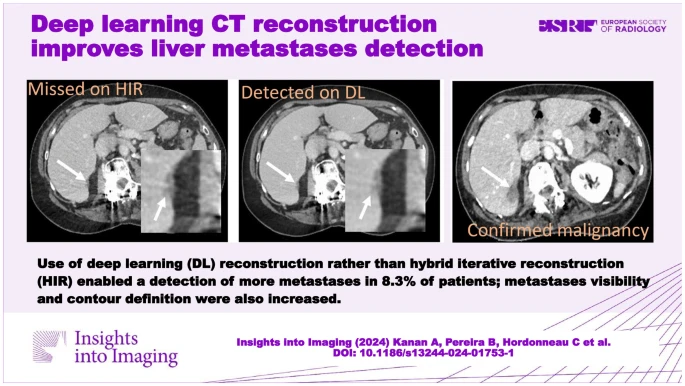
The study hypothesised that the enhanced image quality from DLIR could increase the detection of liver metastases compared to conventional iterative reconstructions. Authors compared the number of detected metastases using three different levels of DLIR and 50%-ASiR-V in 121 patients. The results showed that DLIR-high significantly increased the number of detected lesions for the senior reader, with consensus readings confirming additional detections in ten patients. DLIR-high also received better scores for visibility and contour definition of hepatic metastases from both readers, indicating superior image quality.
Clinical Implications and Limitations
The early and accurate detection of liver metastases is crucial for determining the appropriate management strategies, including surgical resection, ablation, radiotherapy, or systemic therapies. Enhanced detection capabilities, particularly for subcentimeter lesions, can significantly influence treatment planning and outcomes. Despite its potential, DLIR has limitations. High levels of deep learning can sometimes reduce lesion conspicuity, especially for small hepatic and extrahepatic structures. Additionally, while the study showed increased sensitivity for lesion detection, it did not evaluate patient management or outcomes directly.
The study was retrospective and relied on a single CT system and DLIR manufacturer, potentially limiting the generalizability of the findings. The inclusion criterion was based on the final CT report, with histopathological proof of primary cancer, but did not include subgroup analyses based on tumour type. Future studies should address these limitations and evaluate the clinical impact of DLIR on patient management and outcomes.
Deep learning-based reconstructions, specifically TrueFidelity, offer significant improvements in the detection and conspicuity of liver metastases compared to traditional iterative reconstructions like ASiR-V. Using high-strength DLIR statistically increased the number of detected metastases and enhanced image quality, providing a compelling case for its adoption in abdominal CT imaging for oncology. Further research is needed to assess the clinical implications of these findings, but the current evidence supports the integration of deep learning reconstructions to improve early detection and treatment planning for liver metastases.
Source: Insights into Imaging
Image Credit: iStock